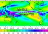
New data reveal an unprecedented depiction of a region of partially molten rock deep within the Earth, which appears to be feeding material in the form of a plume to the surface, where Iceland is located.
The finding, in combination with evidence from previous studies, suggests that these molten regions deep below, near the core-mantle boundary of the Earth, may cause basaltic ocean island chains to form along the surface. Around the Earth’s core-mantle boundary are regions called ultralow-velocity zones (ULVZs), which are characterized by liquid rock with velocities up to 30% lower than surrounding material.
However, depicting ULVZs has been particularly difficult given their extreme depths. ULVZs have been detected below the Polynesian country of Samoa and the Hawaiian islands, yet a clear depiction of their shape has eluded scientists. The proximity of these ULVZs below volcanic island chains has prompted theories suggesting that the giant reservoirs of molten rock feed the mantle plumes that create the islands on Earth’s surface.
Here, Kaiqing Yuan and Barbara Romanowicz used seismic tomography, which constructs an x-ray-like picture of the Earth’s interior using seismic waves, to probe a ULVZ below Iceland.
Based on their results, there appears to be a massive circular blob of partially molten rock, approximately 800 kilometers in diameter and 15 kilometers in height, along the core-mantle boundary, feeding the plume directly below the basaltic island.
The authors note that this ULVZ’s location, shape and large diameter, which is proportionate with the width of the plume higher up in the lower mantle, suggests a close link between the ULVZ and the rising plume above it.
These new data, in combination with the known presence of ULVZs below Samoa and Hawaii, led the authors to propose that a specific class of large ULVZs form at the roots of broad plumes that feed active hotspots.
Reference:
Kaiqing Yuan, Barbara Romanowicz. Seismic evidence for partial melting at the root of major hot spot plumes. Science, 2017; 357 (6349): 393 DOI: 10.1126/science.aan0760
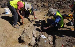
Dr Tim Holland (seated right) assisting volunteers in the excavation of the ribs of Austrosaurus mckillopi in 2015. Credit: Dr Stephen Poropat.
The discovery of new bones belonging to a long-necked sauropod named Austrosaurus mckillopi has been announced by a team of Australian and British palaeontologists.
The bones date from the Early Cretaceous period (104-102 million years ago) and were first discovered in 1932 on Clutha sheep station, northwest of Richmond, Queensland.
However, attempts by palaeontologists to relocate the site during the 1970s and 1990s failed.
Swinburne palaeontologist Dr Stephen Poropat became intrigued by the mystery of the lost site when he studied the bones uncovered in 1932, currently in storage at the Queensland Museum.
“When I realised that the backbones at the museum probably formed a section of a dinosaur’s spine, I hypothesised that more of the skeleton was waiting to be found,” Dr Poropat says.
In 2014, he contacted Dr Tim Holland, former curator of Richmond’s Kronosaurus Korner marine fossil museum, about relocating the site. Dr Holland enlisted the help of Richmond Mayor John Wharton – who grew up on Clutha station – to find the lost site and, hopefully, more of the skeleton.
“When we failed to find the Austrosaurus site at ground level, John jumped into his helicopter,” Dr Holland says.
“From the air we spotted two wooden posts – both of which had toppled over – that had once supported a sign marking the spot.
“John then found fossilised portions of bone embedded in rock nearby. We were blown away.”
Three digs at the site between 2014-2015 uncovered six rib bones, which when placed with the vertebrae found in the early 1930s created a more complete picture of the dinosaur.
“The most exciting realisation was that portions of the ribs were embedded in the rock surrounding the left side of the backbones,” Dr Poropat says.
“This matched the ribs that we found in 2014-2015, five of them from the left side too.
“This means that the carcass of Austrosaurus came to rest on its left side, and it was not shifted much after it died allowing the bones to stay close to a life position.”
Because of its age, Dr Poropat says Austrosaurus might reveal something about the evolution of other sauropods in Australia.
“The sauropods commonly found in the Winton area, south of Richmond, lived five to ten million years after Austrosaurus,” says Dr Poropat. “This means that Austrosaurus could potentially be their close relative or even their direct ancestor.
“Unfortunately the bones are too incomplete and poorly preserved for us to be able to say much with certainty. Nevertheless, we can tell that Austrosaurus was at least distantly related to Winton’s titanosaurs like Diamantinasaurus and Savannasaurus since it shares some features with them.”
Potential for other discoveries
Although the Austrosaurus site is now believed to be exhausted of fossils, the potential for future discoveries of important fossils in the Richmond area is huge.
“Rocks of the right age, deposited in a Cretaceous inland sea known as the Eromanga Sea, are close to the surface all over the Richmond region,” says Dr Holland. “Who knows what else might be waiting to be found? A lucky discovery by a grazier, fossil hunter or tourist out there might be a game-changer.”
The new research on Austrosaurus has been published in Alcheringa, an Australasian Journal of Palaeontology.
Reference:
Stephen F. Poropat et al. Reappraisal of Austrosaurus mckillopi Longman, 1933 from the Allaru Mudstone of Queensland, Australia’s first named Cretaceous sauropod dinosaur, Alcheringa: An Australasian Journal of Palaeontology (2017). DOI: 10.1080/03115518.2017.1334826
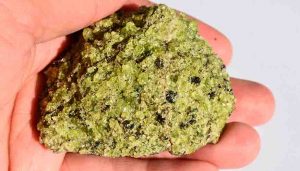
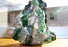
A mantle nodule collected from San Carlos, Arizona, brought to the surface during a deep volcanic eruption about 1 million years ago. Olivine, which is the focus of the LLNL study, is the predominant light green-colored mineral that is present in this rock. Credit: Wyatt Du Frane/LLNL
Hydrogen at elevated temperature creates high electrical conductivity in the Earth’s mantle.
New work by Lawrence Livermore National Laboratory (LLNL) scientists shows the dispersal of water (incorporated as hydrogen in olivine, the most abundant mineral in the upper mantle), could account for high electrical conductivity seen in the asthenosphere (part of the upper mantle just below the lithosphere that is involved in plate tectonic movement). The research appears in Scientific Reports .
The work could lead to a better understanding of present day water distribution in the mantle, which has strong implications for planetary dynamics and evolution. Researchers said such information might provide key evidence as to why Earth is the only known planetary body in our solar system to develop plate tectonics and to retain liquid water oceans on its surface.
“We approached the problem from a different perspective, using new hydrogen diffusion measurements to infer what the contribution of hydrogen would be to electrical conductivity,” said LLNL’s Wyatt Du Frane, the principal investigator on the project. “Our experiments on olivine indicated a larger temperature dependence than previously thought to occur for this phenomenon. The contribution of hydrogen to electrical conductivity, while modest at lower temperatures, becomes quite large at the temperatures expected to occur in the mantle.”
Minerals formed deep in the mantle and transported to the Earth’s surface contain tens to hundreds of parts per million in weight (ppm wt) of water, providing evidence for the presence of dissolved water in the Earth’s interior. Even at these low concentrations, water greatly affects the physico-chemical properties of mantle materials. The diffusion of hydrogen controls the transport of water in the Earth’s upper mantle, but until now was not fully understood for olivine.
Earth’s hydrosphere is a distinctive feature of our planet where massive oceans affect its climate and support its ecosystem. The distribution of water on Earth is not limited to its outermost shell (hydrosphere and hydrated minerals), but extends to great depths within the planet. Downwelling oceanic lithosphere (at subduction zones) and upwelling magmas (at mid ocean ridges, volcanoes and hotspots) are vehicles for transport of H2O between the surface and the Earth’s deep interior.
“The amount of hydrogen required to match geophysical measurements of electrical conductivity inside Earth are in line with the concentrations that are observed in oceanic basalts. This demonstrates that geophysical measurements of electrical conductivity are a promising tool for mapping out water distributions deep inside the Earth,” Du Frane said.
Reference:
Davide Novella et al. Hydrogen self-diffusion in single crystal olivine and electrical conductivity of the Earth’s mantle, Scientific Reports (2017). DOI: 10.1038/s41598-017-05113-6
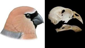
Reconstruction of Pyrrhula crassa (left) and skull (right). Credit: Pau Oliver
Inside the crater of a volcano on Graciosa Island in the Azores archipelago, in the Atlantic Ocean, an international team of researchers has discovered the bones of a new extinct species of songbird, a bullfinch which they have named Pyrrhula crassa. The remains were found in a small cavity through which time ago the lava flowed. This bird disappeared a few hundreds of years ago due to human colonization of the islands and the introduction of invasive species.
Until hundreds of years ago, a species of bullfinch, a small songbird with a very short and robust beak, lived on Graciosa Island in the Azores archipelago. The arrival of humans to this island, however, depleted its population and it ended up going extinct, as was the case with numerous bird species on other islands, such as the Canaries and Madeira.
Now, an international team of scientists, backed by a project led by Josep Antoni Alcover, from the Mediterranean Institute for Advanced Studies (IMEDEA, CSIC-UIB), has discovered the bones of this bullfinch, called Pyrrhula crassa, in a cave located in a 12,000-year-old volcano in the southeast of the island.
“It is the first extinct passerine bird described in the archipelago, and it won’t be the last,” states Alcover, co-author of the study published in Zootaxa which focused on the analysis of beak morphology in order to determine the new species.
Despite there being few known remains of this bird, they are sufficiently distinctive for the scientists to have succeeded in establishing that they belong to a new extinct species of bullfinch.
The new bird, being the largest of its genus according to the size of the skull remains found, recalls due to its flying ability the existing bullfinch from the other Azores island (São Miguel) which is ‘vulnerable’ to extinction because of the expansion of agriculture and the disappearance of laurel forests.
“Its short and wide beak was not just considerably bigger, but also relatively higher than that of the common bullfinch or that from São Miguel, with a very robust configuration reminiscent to an extent of the beak of a small parrot,” asserts the researcher.
Invasions wiped out the birds
These islands were colonized during the 13th century by the Portuguese, although they could have been visited by Vikings over one thousand years ago. Just as has happened on many other islands, such as the Canaries or Madeira, different bird species have disappeared throughout the last millennium due to the arrival of humans along with various invasive species.
Human colonization led to the destruction and burning of the islands’ habitats in which humans started settling, and they impacted on the birds which were part of the indigenous fauna. P. crassa was no exception, finding itself affected until its extinction.
The introduction of invasive plant species has depleted and reduced the area of the laurel forests in which this species of bird lived by up to 3% of its original size. According to the scientists, although remains of P. crassa have only been identified in Graciosa so far, it possibly inhabited other islands of the Azores archipelago.
Reference:
J.C RANDO, H. PIEPER, STORRS L. OLSON, F. PEREIRA, J.A. ALCOVER. A new extinct species of large bullfinch (Aves: Fringillidae: Pyrrhula) from Graciosa Island (Azores, North Atlantic Ocean). Zootaxa, 2017; 4282 (3): 567 DOI: 10.11646/zootaxa.4282.3.9
Note: The above post is reprinted from materials provided by SINC.
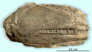
The 26 cm long fossil preserving the right side of the skull of Birgeria americana. Credit: UZH
The most catastrophic mass extinction on Earth took place about 252 million years ago — at the boundary between the Permian and Triassic geological periods. Up to 90 percent of the marine species of that time were annihilated. Worldwide biodiversity then recovered in several phases throughout a period of about five million years. Until now, paleontologists have assumed that the first predators at the top of the food chain did not appear until the Middle Triassic epoch about 247 to 235 million years ago.
Unexpected find of a large predatory fish
Swiss and U.S. American researchers led by the Paleontological Institute and Museum of the University of Zurich have discovered the fossil remains of one of the earliest large-sized predatory fishes of the Triassic period: an approximately 1.8-meter-long primitive bony fish with long jaws and sharp teeth. This fish belongs to a previously unknown species called Birgeria americana. This predator occupied the sea that once covered present-day Nevada and the surrounding states already one million years after the mass extinction.
Triassic “Jaws”
In the United States, almost no vertebrate fossils from the Early Triassic epoch (252 to 247 million years ago) have been scientifically described until now. “The surprising find from Elko County in northeastern Nevada is one of the most completely preserved vertebrate remains from this time period ever discovered in the United States,” emphasizes Carlo Romano, lead author of the study. The fossil in question is a 26-centimeter-long partial skull of a fierce predator, as evidenced by three parallel rows of sharp teeth up to 2 centimeters long along the jaw margins, as well as several smaller teeth inside the mouth.
Birgeria hunted similarly to the extant great white shark: the prey fish were pursued and bitten, then swallowed whole. Species of Birgeria existed worldwide. The most recent discovery is the earliest example of a large-sized Birgeria species, about one and a half times longer than geologically older relatives.
Predators appeared earlier than assumed
According to earlier studies, marine food chains were shortened after the mass extinction event and recovered only slowly and stepwise. In addition, researchers assumed that the ancient equatorial regions were too hot for vertebrates to live during the Early Triassic. Finds such as the newly discovered Birgeria species and the fossils of other vertebrates now show that so-called apex predators (animals at the very top of the food chain) already lived early after the mass extinction. The existence of bony fish close to the equator — where Nevada was located during the Early Triassic — indicates that the temperature of the sea was a maximum of 36°C. The eggs of today’s bony fish can no longer develop normally at constant temperatures above 36°C.
“The vertebrates from Nevada show that previous interpretations of past biotic crises and associated global changes were too simplistic,” Carlo Romano says. Despite the severity of the extinctions of that time and intense climatic changes, the food webs were able to redevelop faster than previously assumed.
Reference:
Carlo Romano, James F. Jenks, Romain Jattiot, Torsten M. Scheyer, Kevin G. Bylund, Hugo Bucher. Marine Early Triassic Actinopterygii from Elko County (Nevada, USA): implications for the Smithian equatorial vertebrate eclipse. Journal of Paleontology, 2017; 1 DOI: 10.1017/jpa.2017.36
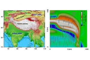
A topographic map of the area around the Tibetan Plateau, left, and the map view of the composite strong and weak Asian plate model, right. The composite plate strength model — with the Asian plate stronger in the west (Tarim Basin) and weaker to the east — results in a topography that is similar to what exists today. Credit: Graphic courtesy of Lin Chen
Geoscientists have long puzzled over the mechanism that created the Tibetan Plateau, but a new study finds that the landform’s history may be controlled primarily by the strength of the tectonic plates whose collision prompted its uplift. Given that the region is one of the most seismically active areas in the world, understanding the plateau’s geologic history could give scientists insight to modern day earthquake activity.
The new findings are published in the journal Nature Communications.
Even from space, the Tibetan Plateau appears huge. The massive highland, formed by the convergence of two continental plates, India and Asia, dwarfs other mountain ranges in height and breadth. Most other mountain ranges appear like narrow scars of raised flesh, while the Himalaya Plateau looks like a broad, asymmetrical scab surrounded by craggy peaks.
“The asymmetric shape and complex subsurface structure of the Tibetan Plateau make its formation one of the most significant outstanding questions in the study of plate tectonics today,” said University of Illinois geology professor and study co-author Lijun Liu.
In the classic model of Tibetan Plateau formation, a fast-moving Indian continental plate collides head-on with the relatively stationary Asian plate about 50 million years ago. The convergence is likely to have caused the Earth’s crust to bunch up into the massive pile known as the Himalaya Mountains and Tibetan Plateau seen today, but this does not explain why the plateau is asymmetrical, Liu Said.
“The Tibetan Plateau is not uniformly wide,” said Lin Chen, the lead author from the Chinese Academy of Sciences. “The western side is very narrow and the eastern side is very broad — something that many past models have failed to explain.”Many of those past models have focused on the surface geology of the actual plateau region, Liu said, but the real story might be found further down, where the Asian and Indian plates meet.
“There is a huge change in topography on the plateau, or the Asian plate, while the landform and moving speed of the Indian plate along the collision zone are essentially the same from west to east,” Liu said. “Why does the Asian plate vary so much?”
To address this question, Liu and his co-authors looked at what happens when tectonic plates made from rocks of different strengths collide. A series of 3-D computational continental collision models were used to test this idea.
“We looked at two scenarios — a weak Asian plate and a strong Asian plate,” said Liu. “We kept the incoming Indian plate strong in both models.”
When the researchers let the models run, they found that a strong Asian plate scenario resulted in a narrow plateau. The weak Asian plate model produced a broad plateau, like what is seen today.
“We then ran a third scenario which is a composite of the strong and weak Asian plate models,” said Liu. “An Asian plate with a strong western side and weak eastern side results in an orientation very similar to what we see today.”
This model, besides predicting the surface topography, also helps explain some of the complex subsurface structure seen using seismic observation techniques.
“It is exciting to see that such a simple model leads to something close to what we observe today,” Liu said. “The location of modern earthquake activity and land movement corresponds to what we predict with the model, as well.”
Reference:
Lin Chen, Fabio A. Capitanio, Lijun Liu, Taras V. Gerya. Crustal rheology controls on the Tibetan plateau formation during India-Asia convergence. Nature Communications, 2017; 8: 15992 DOI: 10.1038/ncomms15992
An article just published in the Proceedings of the Royal Society B describes two remarkably different hydrothermal vent fields discovered in the southern Gulf of California. Despite being relatively close together, these vents host very different animal communities. This finding contradicts a common scientific assumption that neighboring vents will share similar animal communities. Instead, the new paper suggests that local geology and the chemistry of vent fluids are important factors affecting vent communities.
In 2012, scientists from the Monterey Bay Aquarium Research Institute (MBARI) used undersea robots to discover a new hydrothermal vent field along the Alarcón Rise at the southern end of the Gulf of California. Continuing the effort in 2015, they discovered a second, very different vent field in the Pescadero Basin, just 75 kilometers to the north.
Led by MBARI scientists, the research involved scientists from Mexico, Canada, Russia, and Germany. In preparing the recent paper, they analyzed collected organisms and video surveys to determine community composition. They also performed DNA analyses of water samples to identify larvae of vent animals and stable-isotope analysis to assess food supplies at each vent field.
The scientists compared the animals living at the Alarcón and Pescadero Basin vent fields with those found in the Guaymas Basin, 400 kilometers to the north, and on the East Pacific Rise, about 300 kilometers to the south. The researchers found that, despite their close proximity, the Alarcón and Pescadero vent fields support radically different animal communities, sharing only seven out of 61 animal species.
This finding contradicts a common scientific assumption that neighboring habitats will share similar animal communities. Instead, the results suggest that local geology and chemistry of the vent fluids play dominant roles in structuring the animal communities. The findings are relevant to assessing the possible ecological impacts of seafloor mining — scientists must account for the uniqueness of local geology and chemistry and not assume that a common supply of animal larvae will colonize and restore neighboring habitats.
Lead author Shana Goffredi, an MBARI adjunct and associate professor at Occidental College, explained, “Just like human cities, the community that forms in a particular area depends not only on who arrives at that location, but also whether the underlying resources are suitable for their success. Variation in these resources, whether physical or chemical, contributes greatly to the diversity of the region, which is important for community stability.”
Though neighbors, the Alarcón Rise and Pescadero Basin vent fields are geologically very different. The seafloor along the Alarcón Rise is covered in young, fresh lava, and the fluids spewing out of the vents are very hot (up to 360 degrees Celsius) and rich in metal sulfides that form dark, crumbly chimneys known as “black smokers.” Animals at the Alarcón Rise are similar to locations further south (almost 300 kilometers) on the East Pacific Rise.
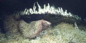
In Pescadero Basin, however, hydrothermal-vent fluids pass through thick layers of seafloor mud. As the hot hydrothermal fluid flows through this mud, it “cooks” organic material, forming methane (natural gas) and oil-like hydrocarbons. The Pescadero Basin vents contain very little sulfide, and the superheated fluids produce giant, light-colored, carbonate chimneys streaked with dark, oily hydrocarbons.
Most of the animals found at the Pescadero vents are worms, and many species are new to science. The dominant tubeworms (genus Oasisia) are not common elsewhere in the Gulf. Surprisingly, two thirds of the Pescadero vent animals are not found at vents to the north and south.
For the last two decades, marine biologists have been trying to document how seafloor animals manage to disperse from one discrete hydrothermal vent habitat to another. The majority of vent animals release microscopic larvae that are carried by ocean currents. If some of these larvae survive long enough to reach another hydrothermal vent, they may settle on the seafloor, grow into adults, and colonize a new vent.
This colonization theory led vent biologists to assume that neighboring vent fields should harbor similar animal communities. However, the new paper shows that larvae from one vent may not successfully colonize a neighboring vent. MBARI researcher Shannon Johnson used high-throughput DNA sequencing to identify larvae collected from the water around the vents. Her results showed that larvae from other sites can reach the Pescadero Basin, but prevailing geological and chemical conditions apparently preclude their settlement and growth there.
The researchers conclude that numerous factors affect the composition of the animal communities found at particular vents. Water depth, geology of the seafloor, temperature and chemistry of the vent fluids, and the ability of larvae from other vents to colonize the site all play roles. Given developing efforts to mine deep-sea hydrothermal vent fields for precious metals, the scientists involved in this research suggest that conservationists and management agencies need to consider a broader range of factors in their efforts to predict the environmental impacts and the resiliency of affected communities.
Reference:
Shana K. Goffredi, Shannon Johnson, Verena Tunnicliffe, David Caress, David Clague, Elva Escobar, Lonny Lundsten, Jennifer B. Paduan, Greg Rouse, Diana L. Salcedo, Luis A. Soto, Ronald Spelz-Madero, Robert Zierenberg, Robert Vrijenhoek. Hydrothermal vent fields discovered in the southern Gulf of California clarify role of habitat in augmenting regional diversity. Proceedings of the Royal Society B: Biological Sciences, 2017; 284 (1859): 20170817 DOI: 10.1098/rspb.2017.0817
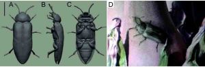
This is a 3-D habitual and environmental reconstructions of Ponomarenkia belmonthensis restored after linedrawing of the holotype and 2-D reconstruction. The plant is Australian cycadophyt Lepidozamia hopei from the Botanical Garden of Jena University. Credit: Evgeny V. Yan/FSU Jena
He’s Australian, around half a centimetre long, fairly nondescript, 300 million years old, and he’s currently causing astonishment among both entomologists and palaeontologists. The discovery of a beetle from the late Permian period, when even the dinosaurs had not yet appeared on the scene, is throwing a completely new light on the earliest developments in this group of insects. The reconstruction and interpretation of the characteristics of Ponomarenkia belmonthensis was achieved by Prof. Dr Rolf Beutel and Dr Evgeny V. Yan of Friedrich Schiller University Jena (Germany). They have published this discovery together with beetle researcher Dr John Lawrence and Australian geologist Dr Robert Beattie in the current issue of the Journal of Systematic Palaeontology. It was Beattie who discovered the only two known fossilised specimens of the beetle in former marshland in Belmont, Australia.
“Beetles, which with nearly 400,000 described species today make up almost one-third of all known organisms, still lived a rather shadowy and cryptic existence in the Permian period,” explains Jena zoologist Beutel. “The fossils known to date have all belonged to an ancestral beetle lineage, with species preferring narrow spaces under bark of coniferous trees. They exhibit a whole series of primitive characteristics, such as wing cases (elytra) that had not yet become completely hardened or a body surface densely covered with small tubercles.”
Earliest form of the modern beetle
In contrast, the species that has now been discovered, assigned to the newly introduced family Ponomarenkiidae, can be identified as a modern beetle, in spite of its remarkable age. Modern characteristics are the antennae resembling a string of beads, antennal grooves, and the unusually narrow abdomen, tapering to a point. What is more, unlike previously known Permian beetles, the wing cases are completely hardened, the body’s surface is largely smooth, and the thoracic segments responsible for locomotion show modern features, notes insect palaeontologist Yan. In addition, it appears that this little beetle had stopped living under tree bark, the habitat favoured by its contemporaries, and had adopted a much more exposed lifestyle on plants. A significant fact is that, due to its unorthodox combination of ancestral and modern characteristics, this genus does not fit in any of the four suborders of beetles that still exist, which is why Yan and Beutel have given it the nickname Bad Boy. “Ponomarenkia belmonthensis shows above all that the first major events of radiation in the evolution of beetles took place before the Permian-Triassic mass extinction,” says Rolf Beutel. Beetles as a whole survived this dramatic event, which saw the acidification of the seas and major volcanic eruptions, considerably better than most other groups of organisms, presumably because of their terrestrial life style and hardened exoskeleton. However, the Bad Boy ran out of luck, as there are no more traces of its existence in the Mesozoic era.
Name honours eminent palaeontologist
The Jena researchers dedicated the genus and family to Moscow palaeontologist Prof. Alexander G. Ponomarenko. He has had a strong influence on beetle palaeontology for decades and supervised Dr Evgeny V. Yan’s doctorate. Yan obtained his doctorate from the Russian Academy of Sciences, spent five years as a postdoc at the Chinese Academy of Sciences in Nanjing, and since June 2016 he has done research at the Institute of Systematic Zoology and Evolutionary Biology with Phyletic Museum of the University of Jena as a guest researcher funded by the Alexander von Humboldt Foundation. It is Yan’s elaborate reconstructions on the computer that have provided the precise insights into Ponomarenkia belmonthensis.
In the first stage, some 40 photographs were taken of the two specimens, which were available as impressions on stone. “With this series of photographs an accurate 2D reconstruction was possible, with which we were able to correct for deformations in the original fossil. This allowed us to get closer to the actual beetle,” explains Dr Yan. Based on precise drawings and with the help of a special computer program that is also used for animation and computer games, a very informative 3D model was created. “The 3D reconstruction also enables us to draw conclusions about the way the beetle moved and lived,” the palaeontologist adds. He has developed this method of visualisation, as well as the analytical process in which he also includes hypothetical ancestors of the beetle, since his arrival in Jena. “We have already been able to apply this process to three newly discovered ancient beetle species,” Prof. Beutel is happy to report. “In this way, we have made significant steps towards deciphering the earliest stages in the evolution of an extremely successful genus of animals.”
Reference:
Evgeny Viktorovich Yan, John Francis Lawrence, Robert Beattie, Rolf Georg Beutel. At the dawn of the great rise: †Ponomarenkia belmonthensis (Insecta: Coleoptera), a remarkable new Late Permian beetle from the Southern Hemisphere. Journal of Systematic Palaeontology, 2017; 1 DOI: 10.1080/14772019.2017.1343259
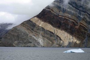
Northeast Greenland National Park is the world’s largest and most northerly national park. It is the largest protected land area in the world. Established in 1974 and expanded to its present size in 1988, it protects 972,001 km2 (375,000 sq mi) of the interior and northeastern coast of Greenland and is bigger than all but twenty-nine countries in the world. It was the first national park to be created in the Kingdom of Denmark and remains Greenland’s only national park.
Stripy cliffs
1,900m-high, stripy cliffs in Segelsällskarpet Fjord. The colorful layers are part of the Eleonore Bay group and are made up of alternating layers of limestone, dolomite, mud rocks and Quartzite’s. Northeast Greenland National Park (Greenlandic: Kalaallit Nunaanni nuna eqqissisimatitaq, Danish: Grønlands Nationalpark) is the world’s largest and most northerly national park. Established in 1974 and expanded to its present size in 1988, it protects 972,001 km2 of the interior and northeast
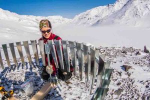
UAF researcher Anna Liljedahl puts up a wind shield around a rain gauge she installed on Jarvis Glacier. Credit: UAF photo by Todd Paris
Small mountain glaciers play a big role in recharging vital aquifers and in keeping rivers flowing during the winter, according to a new study published in Geophysical Research Letters, a journal of the American Geophysical Union.
The study also suggests that the accelerated melting of mountain glaciers in recent decades may explain a phenomenon that has long puzzled scientists — why Arctic and sub-Arctic rivers have increased their water flow during the winter even without a correlative increase in rain or snowfall.
“I think that mountain glaciers in the Arctic and sub-Arctic have really been underappreciated as a source of water to the landscape,” said Anna Liljedahl, the lead author and an associate professor at the University of Alaska Fairbanks’ Water and Environmental Research Center.
Liljedahl and her co-authors at the U.S. Geological Survey and U.S. Army Corps of Engineers’ Cold Regions Research and Engineering Laboratory studied a watershed in a semidry climate in the eastern Alaska Range. The team looked at how meltwater from two small mountain glaciers flowed through the system and influenced the mountain streams, rivers and groundwater all year long.
Through extensive field measurements, the team found that Jarvis and Gulkana glaciers contributed 15 percent to 66 percent of the annual flow in mountain streams that drain the glaciers’ meltwater into the Delta River. Yet when the team compared the volume of water at an upper site and another site about 35 miles downstream on one of the major mountain streams, they found that the stream lost half its water to an aquifer storing groundwater.
“These headwater streams, coming off the mountains and into the lowland, are like the water line to your house peppered with holes, half of the water disappearing into the ground and recharging your neighbor’s house well instead of it all reaching your kitchen faucet,” said Liljedahl.
Liljedahl said the recharge of the aquifers is important because they don’t freeze during the winter and are the only source of water to the rivers during this time. In the town of Delta Junction, which sits adjacent to the Delta River far downstream from the Jarvis and Gulkana glaciers, the water table drops more than 33 feet each winter as the aquifers drain. The water leaks into the Tanana River, which the Delta River also feeds.
Liljedahl said this process has probably continued for thousands of years. Yet recent temperature gains in climate may be accelerating the glaciers’ melting and introducing more meltwater into aquifers and then the rivers.
“The winter discharge of the Tanana River has increased since the record keeping began in the ’70s, but there are no increasing trends in precipitation,” she said. “Glacier coverage has, on the other hand, decreased by 12 percent, and that is more than plenty of additional water to explain the increase in river base flow. In fact, about five times more.”
But it may not be long before this process ends all together, she said. The glaciers are disappearing or shrinking to very high elevations where colder temperatures slow melting. As glacier melt decreases, so may the stream flow if there is not enough water to both feed both the aquifer and the stream.
Co-author Shad O’Neel, a researcher with the U.S. Geological Survey’s Alaska Science Center, said this study shows another way that glaciers are connected to the ecosystem and to humans in the Arctic.
“Although the traditional focus of glaciology has been on sea level rise, we are rapidly discovering that the small mountain glaciers may have large impacts on human populations,” he said. “Across the globe, the mountain glaciers influence ecosystem processes like stream flow, nutrient delivery and primary production in the ocean. Human implications range from drinking and agricultural water supplies to recreation and tourism. ”
Michel Baraer, professor at École de Technologie Supérieure in Quebec, Canada, said the study only marks the beginning of research on links between glaciers and groundwater.
“No doubt that it will be the precursor of new research in that field,” he said.
Liljedahl said she is currently researching the role of mountain glacier melt in the water cycle in other semiarid landscapes, such as those in the Russian and Canadian Arctic. If the process holds true in these places, she said, then tiny mountain glaciers may be helping power watersheds throughout large portions of the Arctic.
“I think people have assumed that these tiny glaciers are not important because they’re tiny, but we’re in a climate where we have very little precipitation,” she said. “Any additional water can make a big splash.”
Reference:
A. K. Liljedahl, A. Gädeke, S. O’Neel, T. A. Gatesman, T. A. Douglas. Glacierized headwater streams as aquifer recharge corridors, subarctic Alaska. Geophysical Research Letters, 2017; DOI: 10.1002/2017GL073834
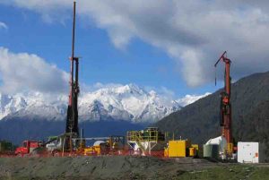
The Deep Fault Drilling Project borehole. Credit: John Townend, Victoria University, NZ
Analysis of natural sparkling mineral water has given scientists valuable clues on how to locate hot water springs — potential sources of sustainable, clean energy.
Studies at naturally carbonated springs have shown how oxygen in the water comes to have a distinctive chemical fingerprint.
Research showed that this fingerprint is influenced by the presence of carbon dioxide gas — and not by heat from below Earth’s surface, as was previously thought.
The finding may help scientists narrow their search for sites where geothermal energy — heat generated and stored in Earth — could be sustainably recovered.
Scientists from the University of Edinburgh analysed water from naturally carbonated springs in Daylesford, Australia, and Pah Tempe in Utah, US.
The team used computers to model the interactions between the water and surrounding rocks, based on measurements of water samples from the sites. Their findings eliminated the possibility that minerals from the rocks affected the oxygen in the water. Instead, they showed that CO2 gas must be influencing the oxygen’s composition.
The study, published in Applied Geochemistry, was supported by the UK Engineering and Physical Sciences Research Council and the Australian research organisation CO2CRC.
R?ta Karolyt?, of the School of GeoSciences, who led the study, said: “The oxygen fingerprint of spring waters has long been used to estimate the depth of the water’s source. Our new finding, that the mixing of natural CO2 with water changes its oxygen fingerprint, means that many sparkling spring waters previously thought to be originating from very deep in Earth’s crust actually only have this fingerprint because of mixing with CO2.”
Dr Stuart Gilfillan, of the School of GeoSciences, who co-ordinated the study, said “This finding changes how we can use the oxygen fingerprints of natural spring waters to identify potential geothermal resources. Estimates of how much heat a sparkling water spring has been exposed to should take into account the effect of CO2.”
Reference:
Rūta Karolytė, Sascha Serno, Gareth Johnson, Stuart M.V. Gilfillan. The influence of oxygen isotope exchange between CO 2 and H 2 O in natural CO 2 -rich spring waters: Implications for geothermometry. Applied Geochemistry, 2017; 84: 173 DOI: 10.1016/j.apgeochem.2017.06.012
Researchers have found that the formation and breakup of supercontinents over hundreds of millions of years controls volcanic carbon emissions. The results, reported in the journal Science, could lead to a reinterpretation of how the carbon cycle has evolved over Earth’s history, and how this has impacted the evolution of Earth’s habitability.
The researchers, from the University of Cambridge, used existing measurements of carbon and helium from more than 80 volcanoes around the world in order to determine its origin. Carbon and helium coming out of volcanoes can either come from deep within the Earth or be recycled near the surface, and measuring the chemical fingerprint of these elements can pinpoint their source. When the team analysed the data, they found that most of the carbon coming out of volcanoes is recycled near the surface, in contrast with earlier assumptions that the carbon came from deep in the Earth’s interior. “This is an essential piece of geological carbon cycle puzzle,” said Dr Marie Edmonds, the senior author of the study.
Over millions of years, carbon cycles back and forth between Earth’s deep interior and its surface. Carbon is removed from the surface from processes such as the formation of limestone and the burial and decay of plants and animals, which allows atmospheric oxygen to grow at the surface. Volcanoes are one way that carbon is returned to the surface, although the amount they produce is less than a hundredth of the amount of carbon emissions caused by human activity. Today, the majority of carbon from volcanoes is recycled near the surface, but it is unlikely that this was always the case.
Volcanoes form along large island or continental arcs where tectonic plates collide and one plate slides under the other, such as the Aleutian Islands between Alaska and Russia, the Andes of South America, the volcanoes throughout Italy, and the Mariana Islands in the western Pacific. These volcanoes have different chemical fingerprints: the ‘island arc’ volcanoes emit less carbon which comes from deep in the mantle, while the ‘continental arc’ volcanoes emit far more carbon which comes from closer to the surface.
Over hundreds of millions of years, the Earth has cycled between periods of continents coming together and breaking apart. During periods when continents come together, volcanic activity was dominated by island arc volcanoes; and when continents break apart, continental volcano arcs dominate. This back and forth changes the chemical fingerprint of carbon coming to Earth’s surface systematically over geological time, and can be measured through the different isotopes of carbon and helium.
Variations in the isotope ratio, or chemical fingerprint, of carbon are commonly measured in limestone. Researchers had previously thought that the only thing that could change the carbon fingerprint in limestone was the production of atmospheric oxygen. As such, the carbon isotope fingerprint in limestone was used to interpret the evolution of habitability of Earth’s surface. The results of the Cambridge team suggest that volcanoes played a larger role in the carbon cycle than had previously been understood, and that earlier assumptions need to be reconsidered.
“This makes us fundamentally re-evaluate the evolution of the carbon cycle,” said Edmonds. “Our results suggest that the limestone record must be completely reinterpreted if the volcanic carbon coming to the surface can change its carbon isotope composition.”
A great example of this is in the Cretaceous Period, 144 to 65 million years ago. During this time period there was a major increase in the carbon isotope ratio found in limestone, which has been interpreted as an increase in atmospheric oxygen concentration. This increase in atmospheric oxygen was causally linked to the proliferation of mammals in the late Cretaceous. However, the results of the Cambridge team suggest that the increase in the carbon isotope ratio in the limestones could be almost entirely due to changes in the types of volcanoes at the surface.
“The link between oxygen levels and the burial of organic material allowed life on Earth as we know it to evolve, but our geological record of this link needs to be re-evaluated,” said co-author Dr Alexandra Turchyn, also from the Department of Earth Sciences.
Reference:
Emily Mason et al. Remobilization of crustal carbon may dominate volcanic arc emissions, Science (2017). DOI: 10.1126/science.aan5049
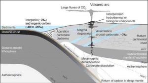
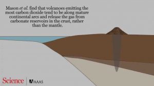
This image depicts results by Mason et al. This material relates to a paper that appeared in the July 21, 2017 issue of Science, published by AAAS. The paper, by E. Mason at University of Cambridge in Cambridge, UK, and colleagues was titled, ‘Remobilization of crustal carbon may dominate volcanic arc emissions.’ Credit: Carla Schaffer / AAA
A new analysis suggests that much of the carbon released from volcanic arcs, chains of volcanoes that arise along the tectonic plates of a subduction zone, comes from remobilizing limestone reservoirs in the Earth’s crust. Previous research suggested carbon was sourced from the mantle as a result of the subduction process.
The discovery ultimately impacts the amount of organic carbon scientists believe was buried in the past. Carbon cycling between surface reservoirs and the mantle over geologic history is important because the imbalance greatly influences the amount of total carbon at Earth’s surface. However, the source for carbon from volcanic arc outgassing remained uncertain.
Emily Mason and colleagues compiled a global data set of carbon and helium isotopes to determine the origin of the carbon. The data reveal that many volcanic arcs mobilize carbon from large, crustal carbonate platforms — particularly in Italy, the Central American Volcanic Arc, Indonesia, and Papua New Guinea.
In contrast, arcs located in the northern Pacific, such as Japan and Kuril-Kamchatka, release carbon dioxide with an isotope signature indicative of a mantle source.
The recognition of a large amount of crustal carbon in the overall carbon isotope signature requires, from a mass balance consideration, downward revision of how much organic carbon was buried in the past.
Reference:
Emily Mason, Marie Edmonds, Alexandra V. Turchyn. Remobilization of crustal carbon may dominate volcanic arc emissions. Science, 2017; 357 (6348): 290 DOI: 10.1126/science.aan5049
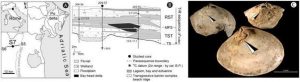
Location map, cross-section, and images of parasitized Abra segmentum valves. A-Location map of investigated Po coastal plain sector, Italy B-Cross section illustrating core samples. C-Photomicrographs of A. segmentum with trematode-induced pits. Credit: Scientific Reports
In 2014, a team of researchers led by a paleobiologist from the University of Missouri found that clams from the Holocene Epoch (that began 11,700 years ago) contained clues about how sea level rise due to climate change could foreshadow a rise in parasitic trematodes, or flatworms. The team cautioned that the rise could lead to outbreaks in human infections if left unchecked. Now, an international team from Mizzou and the Universities of Bologna and Florida has found that rising seas could be detrimental to human health on a much shorter time scale. Findings from their study in northern Italy suggest that parasitic infections could increase in the next century, if history repeats itself.
Trematodes are internal parasites that affect mollusks and other invertebrates inhabiting estuarine environments, which are the coastal bodies of brackish water connecting rivers to the open sea. John Huntley, assistant professor of geological sciences in the MU College of Arts and Science, studied the prehistoric clams as a senior visiting fellow for the Institute for Advanced Studies at the University of Bologna, Italy. With core samples taken from the Po River plain in Italy, the team found traces made by trematodes on the shells of the clams disclosing the connections between the ancient clams and climate change.
“The forecasts of increasing global temperatures and sea level rise have led to major concerns about the response of parasites to climate change,” Huntley said. “Italy has a robust environmental monitoring program, so there was a wealth of information to examine.”
Ancient trematodes had soft bodies; therefore, they didn’t leave body fossils. However, infected clams developed oval-shaped pits around the parasite in the attempt to keep it out, and it’s the prevalence of those pits and their makeup that provide clues as to what happened during different eras in time.
Using 61 samples collected from a drill core obtained by the Italian government for geological research, the scientists examined trematode traces and matched the information to existing records measuring sea level and salinity rises through the ages.
“We found that pulses in sea-level rise occurred on the scale of hundreds of years, and that correlated to rises in parasitic trematodes in the core samples,” Huntley said. “What concerns me is that these rises are going to continue to happen and perhaps at accelerated rates. This poses grave concerns for public health and ecosystem services. These processes could increase parasitism in not only estuarine systems but also in freshwater settings. Such habitats are home to the snail hosts of blood flukes, which infect and kill a million or more people globally each year. What’s scary is it could potentially affect the generations of our kids or grandkids.”
Huntley and his team think that the discoveries they continue to make about impending climate change could provide a good road map for conservationists and those making decisions about marine environments worldwide.
Reference:
Daniele Scarponi, Michele Azzarone, Michał Kowalewski, John Warren Huntley. Surges in trematode prevalence linked to centennial-scale flooding events in the Adriatic. Scientific Reports, 2017; 7 (1) DOI: 10.1038/s41598-017-05979-6
Scientists are working hard to determine the how, why and when of earthquakes, but getting answers is a complex team effort, says a Victoria University of Wellington geophysicist.
It’s 30 years since John Townend recalls first experiencing a big earthquake—the magnitude 6.5 Edgecumbe earthquake, which struck in March 1987 less than 100 kilometres from his high school in Rotorua.
The Professor of Geophysics and Head of Victoria’s School of Geography, Environment and Earth Sciences has been studying the physics of earthquakes ever since.
The last few years have seen Professor Townend called on many times for his expertise, most recently following the magnitude 7.8 Kaikoura earthquake in November 2016 when he provided expert commentary in the media explaining what had happened and what was likely to come.
As he will discuss in his upcoming inaugural professorial lecture, recent observations of large and small earthquakes in New Zealand and worldwide have hugely expanded geoscientific knowledge.
“The basic problem is that the big earthquakes we’re concerned about as a society and which we most want to understand occur infrequently, whereas the little ones don’t have much effect but occur often enough to test and refine our ideas,” says Professor Townend.
“To really understand how the earthquake machine operates, we need to combine measurements and theory spanning many orders of magnitude. Working out what is happening is a community effort—many different types of observation and scientific expertise are required.”
In his lecture, Professor Townend will discuss what faults look like at different scales, and what we do and don’t know about how earthquakes are generated and how they interact.
“Projects like the Deep Fault Drilling Project, which drilled nearly 900 metres into the South Island’s Alpine Fault, are helping us understand the health of a major fault—the temperatures, pressures, and stresses it’s subjected to—before an expected large earthquake occurs,” he says.
The Alpine Fault produces earthquakes of around magnitude 8 approximately every 300 years and last ruptured in 1717 AD, says Professor Townend, so understanding what processes control the rupture and reloading of the fault is an urgent scientific and societal challenge.
Meanwhile, data collected during and after the Kaikoura earthquake reveal to seismologists just how finely balanced some faults are.
“The Kaikoura earthquake triggered earthquakes and deep slow slip extending hundreds of kilometres along the Hikurangi subduction zone, below the east coast of the North Island. It’s important that we improve our understanding of what factors make different faults susceptible to slip and what factors control the sizes of the earthquakes that result,” says Professor Townend.
“In a country as geologically young and complex as Aotearoa, earthquakes provide a regular and sometimes devastating reminder that the Earth is in motion.”
Examining conditions within the Earth’s interior is crucial not only to give us a window back to Earth’s history but also to understand the current environment and its future.
This study, published in Nature Communications, offers an explanation of carbon’s descent into the deep Earth. “The stability regions of carbonates are key to understanding the deep carbon cycle and the role of the deep Earth in the global carbon cycle.” says Leonid Dubrovinsky, from the University of Bayreuth.
This is where the ESRF, the European Synchrotron in Grenoble, France. comes in. “The intense X-rays from the ESRF allow us to access the extreme conditions within the entire Earth’s mantle.” underlines Valerio Cerantola, lead author, former PhD student at the University of Bayreuth and now postdoctoral scientist at the ESRF.
In the last century, the rapid increase in the amount of CO2 in the atmosphere together with the observed climate change have increasingly focused scientists’ attention on the carbon cycle and its evolution at the Earth’s surface. The carbon cycle also extends below the surface: recent estimations locate up to 90% of the Earth’s carbon budget in the Earth’s mantle and core. Due to the dynamic nature of tectonic plate movements, convection and subduction, there is a constant recycling of carbon between the Earth’s surface and its deep interior.
In this study, the research team focused on carbonate phases, which are one of the main carbon-bearing minerals in the deep mantle. Carbonates are a group of minerals that contain the carbonate ion (CO32-) and a metal, such as iron or magnesium. The scientists studied the behaviour of a pure iron carbonate, FeCO3 (called siderite), at extreme temperature and pressure conditions covering the entire Earth’s mantle, meaning over 2500 K and 100 GPa, which corresponds to roughly one million times the atmospheric pressure.
“This iron carbonate is of particular interest because of its stability at lower mantle conditions due to spin transition. Moreover the crystal chemistry of the high-pressure carbonates is dramatically different from that at ambient conditions.” explains Elena Bykova, from the University of Bayreuth.
In order to study the stability of FeCO3, the research team performed high pressure and high temperature experiments at three ESRF beamlines: ID27, ID18 and ID09a (now ID15b). “The combination of the multiple techniques gave us unique datasets that ultimately allowed us to uncover new C-carriers inside the deep Earth and show the mechanism behind their formation” says Cerantola. One experimental run was carried out at beamline 13ID-D at APS.
Upon heating FeCO3 to Earth geotherm temperatures at pressures up to about 50 GPa, FeCO3 partially dissociated and formed various iron oxides. At higher pressures, above ~75 GPa, the scientists discovered two new compounds — tetrairon (III) orthocarbonate, Fe43+C3O12, and diiron (II) diiron (III) tetracarbonate, Fe22+Fe23+C4O13.
“There were some theoretical predictions, but so far experimental information about structures of high pressure carbonates have been too limited (and indeed controversial) to speculate about carbonate crystal chemistry. Our data show that while crystal structure of Fe22+Fe23+C4O13 could be found in silicates, no analogues of Fe43+C3O12 are found in nature.” underlines Bykova.
They also found out that one phase, the tetracarbonate Fe4C4O13, shows unprecedented structural stability and keeps its structure even at pressures along the entire geotherm to depths of at least 2500 km, which is close to the boundary between the mantle and the core. It thus demonstrated that self-oxidation-reduction reactions can preserve carbonates in the Earth’s lower mantle. “The study shows the importance of oxidation and reduction (redox) reactions in the deep carbon cycle, which are inevitably linked to other volatile cycles such as oxygen.” underlines Catherine McCammon, from the University of Bayreuth.
Reference:
Valerio Cerantola, Elena Bykova, Ilya Kupenko, Marco Merlini, Leyla Ismailova, Catherine McCammon, Maxim Bykov, Alexandr I. Chumakov, Sylvain Petitgirard, Innokenty Kantor, Volodymyr Svitlyk, Jeroen Jacobs, Michael Hanfland, Mohamed Mezouar, Clemens Prescher, Rudolf Rüffer, Vitali B. Prakapenka, Leonid Dubrovinsky. Stability of iron-bearing carbonates in the deep Earth’s interior. Nature Communications, 2017; 8: 15960 DOI: 10.1038/NCOMMS15960
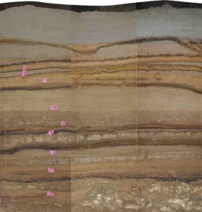
The stratigraphy of the sea cave in Sumatra excavated by scientists from the Earth Observatory of Singapore, Rutgers and other institutions. The lighter bands are sand deposited by tsunamis over a period of 5,000 years; the darker bands are organic material. Credit: Earth Observatory of Singapore
An international team of scientists digging in a sea cave in Indonesia has discovered the world’s most pristine record of tsunamis, a 5,000-year-old sedimentary snapshot that reveals for the first time how little is known about when earthquakes trigger massive waves.
“The devastating 2004 Indian Ocean tsunami caught millions of coastal residents and the scientific community off-guard,” says co-author Benjamin Horton, a professor in the Department of Marine and Coastal Sciences at Rutgers University-New Brunswick. “Our geological record from a cave illustrates that we still cannot predict when the next earthquake will happen.”
“Tsunamis are not evenly spaced through time,” says Charles Rubin, the study’s lead author and a professor at the Earth Observatory of Singapore, part of Nanyang Technological University. “Our findings present a worrying picture of highly erratic tsunami recurrence. There can be long periods between tsunamis, but you can also get major tsunamis that are separated by just a few decades.”
The discovery, reported in the current issue of Nature Communications, logs a number of firsts: the first record of ancient tsunami activity found in a sea cave; the first record for such a long time period in the Indian Ocean; and the most pristine record of tsunamis anywhere in the world.
The discovery was made in a sea cave on the west coast of Sumatra in Indonesia, just south of the city of Banda Aceh, which was devastated by the tsunami of December 2004. The stratigraphic record reveals successive layers of sand, bat droppings and other debris laid down by tsunamis between 7,900 and 2,900 years ago. The stratigraphy since 2,900 years ago was washed away by the 2004 tsunami.
The L-shaped cave had a rim of rocks at the entrance that trapped successive layers of sand inside. The researchers dug six trenches and analyzed the alternating layers of sand and debris using radio carbon dating. The researchers define “pristine” as stratigraphic layers that are distinct and easy to read. “You have a layer of sand and a layer of organic material that includes bat droppings, so simply it is a layer of sand and a layer of bat crap, and so on, going back for 5,000 years,” Horton says.
The record indicates that 11 tsunamis were generated during that period by earthquakes along the Sunda Megathrust, the 3,300-mile-long fault running from Myanmar to Sumatra in the Indian Ocean. The researchers found there were two tsunami-free millennia during the 5,000 years, and one century in which four tsunamis struck the coast. In general, the scientists report, smaller tsunamis occur relatively close together, followed by long dormant periods, followed by great quakes and tsunamis, such as the one that struck in 2004.
Rubin, Horton and their colleagues were studying the seismic history of the Sunda Megathrust, which was responsible for the 2004 earthquake that triggered the disastrous tsunami. They were looking for places to take core samples that would give them a good stratigraphy.
This involves looking for what Horton calls “depositional places” – coastal plains, coastal lake bottoms, any place to plunge a hollow metal cylinder six or seven meters down and produce a readable sample. But for various reasons, there was no site along the southwest coast of Sumatra that would do the job. But Patrick Daly, an archaeologist at EOS who had been working on a dig in the coastal cave, told Rubin and Horton about it and suggested it might be the place they were looking for.
Looking for tsunami records in a sea cave was not something that would have occurred to Horton, and he says Daly’s professional generosity – archaeologists are careful about who gets near their digs – and his own and Rubin’s openness to insights from other disciplines made the research possible. Horton says this paper may be the most important in his career for another reason.
“A lot of (the research) I’ve done is incremental,” he says. “I have a hypothesis, and I do deductive science to test the hypothesis. But this is really original, and original stuff doesn’t happen all that often.”
Reference:
Highly variable recurrence of tsunamis in the 7,400 years before the 2004 Indian Ocean tsunami. Nature Communications (2017). DOI: 10.1038/NCOMMS16019
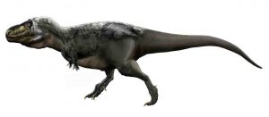
Illustration of the tyrannosaurid Tyrannosaurus rex with minimal feathers based on phylogenetic bracketing. Credit: Durbed/Wikipedia
No other animal on land is faster than a cheetah — the elephant is indeed larger, but slower. For small to medium-sized animals, larger also means faster, but for really large animals, when it comes to speed, everything goes downhill again. For the first time, it is now possible to describe how this parabola-like relationship between body size and speed comes about. A research team under the direction of the German Centre for Integrative Biodiversity Research (iDiv) and the Friedrich Schiller University Jena (Germany) have managed to do so thanks to a new mathematical model, and also published their findings in the journal Nature Ecology and Evolution.
A beetle is slower than a mouse, which is slower than a rabbit, which is slower than a cheetah… which is slower than an elephant? No! No other animal on land is faster than a cheetah — the elephant is indeed larger, but slower. For small to medium-sized animals, larger also means faster, but for really large animals, when it comes to speed, everything goes downhill again. For the first time, it is now possible to describe how this parabola-like relationship between body size and speed comes about.
The model is amazingly simple: The only information that it must be ‘fed’ with is the weight of a particular animal as well as the medium it moves in, so either land, air or water. On this basis alone, it calculates the maximum speed that an animal can reach with almost 90% accuracy. “The best feature of our model is that it is universally applicable,” says the lead author of the study, Myriam Hirt of the iDiv research centre and the University of Jena. “It can be performed for all body sizes of animals, from mites to blue whales, with all means of locomotion, from running and swimming to flying, and can be applied in all habitats.” Moreover, the model is by no means limited to animal species that currently exist, but can be applied equally well to extinct species.
Tyrannosaurus reached a speed of only 17 miles/hour
“To test whether we can use our model to calculate the maximum speed of animals that are already extinct, we have applied it to dinosaur species, whose speed has up to now been simulated using highly complex biomechanical processes,” explains Hirt. The result is that the simple model delivered results for Triceratops, Tyrannosaurus, Brachiosaurus and others that matched those from complex simulations — and were not exactly record-breaking for Tyrannosaurus, who reached a speed of only 27 km/h (17 mi/h). “This means that in future, our model will enable us to estimate, in a very simple way, how fast other extinct animals were able to run,” says the scientist.
Mass has to overcome inertia
Two assumptions are the basis of the model. The first assumption is related on the fact that animals reach their maximum speeds during comparatively short sprints, and not while running over long distances. Unlike running over longer distances, where the body constantly resupplies the muscles with energy (aerobic metabolism), sprinting uses energy that is stored in the muscles themselves but which is exhausted relatively quickly (anaerobic metabolism). It seems logical enough: the larger the animal, the more muscle it has — and thus the faster it can sprint. However, Newton’s laws of motion also apply in the animal kingdom, we know mass has to overcome inertia, and so a five-tonne African elephant simply cannot start moving as quickly as a 2.5-gramme Etruscan shrew. By the time large animals such as the elephant get up to full speed while sprinting, their rapidly available energy reserves also soon run out. Taken together, these two assumptions result in the previously mentioned curve: A beetle is slower than a mouse, which is slower than a rabbit, which is slower than a cheetah — which is faster than an elephant.
Reference:
Myriam R. Hirt, Walter Jetz, Björn C. Rall, Ulrich Brose. A general scaling law reveals why the largest animals are not the fastest. Nature Ecology & Evolution, 2017; DOI: 10.1038/s41559-017-0241-4
For three years, Tara Djokic, a Ph.D. student at the University of New South Wales Sydney, scoured the forbidding landscape of the Pilbara region of Western Australia looking for clues to how ancient microbes could have produced the abundant stromatolites that were discovered there in the 1970s.
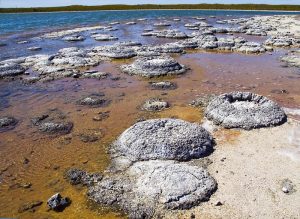
Stromatolites are round, multilayered mineral structures that range from the size of golf balls to weather balloons and represent the oldest evidence that there were living organisms on Earth 3.5 billion years ago.
Scientists who believed life began in the ocean thought these mineral formations had formed in shallow, salty seawater, just like living stromatolites in the World Heritage-listed area of Shark Bay, which is a two-day drive from the Pilbara.
But what Djokic discovered amid the strangling heat and blood-red rocks of the region was evidence that the stromatolites had not formed in salt water but instead in conditions more like the hot springs of Yellowstone.
The discovery pushed back the time for the emergence of microbial life on land by 580 million years and also bolstered a paradigm-shifting hypothesis laid out by UC Santa Cruz astrobiologists David Deamer and Bruce Damer: that life began, not in the sea, but on land.
Djokic’s discovery — together with research carried out by the UC Santa Cruz team, Djokic, and Martin Van Kranendonk, director of the Australian Centre for Astrobiology — is described in an eight-page cover story in the August issue of Scientific American.
“What she (Djokic) showed was that the oldest fossil evidence for life was in fresh water,” said Deamer, a lanky 78-year-old who explored the region with Djokic, Damer, and Van Kranendonk in 2015. “It’s a logical continuation to life beginning in a freshwater environment.”
The model for life beginning on land rather than in the sea could not only reshape our idea about the origin of life and where else it might be, but even change the way we view ourselves.
The right conditions for life
For four decades, ever since the research vessel Alvin discovered deep-sea hydrothermal vents that were habitats for specialized bacteria and worms that looked like something out of a science-fiction novel, scientists have theorized that these mineral- and gas-pumping vents were just what was needed for life to begin.
But Deamer, who describes himself as a scientist who loves playing with new ideas, thought the theory had flaws. For instance, molecules essential for the origin of life would be dispersed too quickly into a vast ocean, he thought, and salty seawater would inhibit some of the processes he knew are necessary for life to begin.
Deamer had spent the early part of his career studying the biophysics of membranes composed of soap-like molecules that form the microscopic boundaries of all living cells. Later, given a piece of the Murchison meteorite that had landed in Australia in 1969, Deamer found that the space rock also contained soap-like molecules nearly 5 billion years old that could form stable membranes. Still later, he demonstrated that membranes helped small molecules join together to form longer information-carrying molecules called polymers.
Trekking to volcanoes from Russia to Iceland and hiking through the Pilbara desert, Deamer and his colleagues observed volcanic activity that suggested the idea that hot springs provided the right environment for the beginning of life. Deamer even built a machine that simulated the heat, acidity, and wet-and-dry cycles of hot springs and installed it in his lab on the UC Santa Cruz campus.
“I think, every once in awhile, you have to be brave enough and bold enough to try new ideas,” Deamer said. “Of course, some of my colleagues think even ‘foolish enough.’ But that’s the chance you take.”
Rethinking the timeline
In Deamer’s vision, ancient Earth consisted of a huge ocean spotted with volcanic land masses. Rain would fall on the land, creating pools of fresh water that would be heated by geothermal energy and then cooled by runoff. Some of the key building blocks of life, created during the formation of our solar system, would have fallen to Earth and gathered in these pools, becoming concentrated enough to form more complex organic compounds.
The edges of the pools would go through periods of wetting and drying as water levels rose and fell. During these periods of wet and dry, lipid membranes would first help stitch together the organic compounds called polymers and then form compartments that encapsulated different sets of these polymers. The membranes would act like incubators for the functions of life.
Deamer and his team believe the first life emerged from the natural production of vast numbers of such membrane-encased “protocells.”
While there is still debate about whether life began on land or in the sea, the discovery of ancient microbial fossils in a place like the Pilbara shows that these geothermal areas — full of energy and rich in the minerals necessary for life — harbored living microorganisms far earlier than believed.
The search for life on other planets
According to Deamer and his colleagues, this discovery and their hot-springs-origins model also have implications for the search for life on other planets. If life began on land, then Mars, which was found to have a 3.65-billion-year-old hot spring deposits similar to those found in the Pilbara region of Australia, might be a good place to look.
For Damer, the new “end-to-end hypothesis” of how life began on land offers something else: that the origin of life was not just a simple story of individual, competing cells. Rather that a plausible new vision of life’s start could be a communal unit of protocells that survived and evolved through collaboration and sharing of innovation rather than strict competition.
“That,” he said, “is a fundamental shift that might impact how we think of our world, ourselves, and our future: as dependent on collaboration as much as being driven by competition.”
Sitting in his fourth-floor office on campus, Deamer smiled as he recounted the letter Charles Darwin wrote to a friend in 1871, which speculated that life might have begun in “some warm little pond.”
That’s not far off the mark, Deamer said, “except we call ours ‘hot little puddles.'”
The Ijen volcano complex is a group of stratovolcanoes in the Bondowoso Regency of East Java, Indonesia. It is inside a larger caldera Ijen, which is about 20 kilometers wide. The Gunung Merapi stratovolcano is the highest point of that complex. The name “Gunung Merapi” means “mountain of fire” in the Indonesian language (api being “fire”); Mount Merapi in central Java and Marapi in Sumatra have the same etymology.
“This blue glow—unusual for a volcano—isn’t, of course, lava, as unfortunately can be read on many websites,” Grunewald told National Geographic in an email about Kawah Ijen, a volcano on the island of Java.
The glow is actually the light from the combustion of sulfuric gases, Grunewald explained.