Fossils preserved in Dominican amber reveal a new family, genus and species of microinvertebrate from the mid-Tertiary period, a discovery that shows unique lineages of the tiny creatures were living 30 million years ago. Credit: Provided by George Poinar Jr.
Fossils preserved in Dominican amber reveal a new family, genus and species of microinvertebrate from the mid-Tertiary period, a discovery that shows unique lineages of the tiny creatures were living 30 million years ago.
The findings by George Poinar Jr. of the Oregon State University College of Science give a rare look at a heretofore unknown clade of invertebrates, along with their fungal food source and other animals that lived in their habitat.
Poinar, an international expert in using plant and animal life forms preserved in amber to learn more about the biology and ecology of the distant past, informally calls the new animals “mold pigs” for their resemblance to swine, and their diet. Scientifically, they are Sialomorpha dominicana, from the Greek words for fat hog (sialos) and shape (morphe).
Invertebrate means not having a backbone, and invertebrates account for roughly 95 percent of animal species.
“Every now and then we’ll find small, fragile, previously unknown fossil invertebrates in specialized habitats,” Poinar said. “And occasionally, as in the present case, a fragment of the original habitat from millions of years ago is preserved too. The mold pigs can’t be placed in any group of currently existing invertebrates — they share characteristics with both tardigrades, sometimes referred to as water bears or moss pigs, and mites but clearly belong to neither group.”
The several hundred individual fossils preserved in the amber shared warm, moist surroundings with pseudoscorpions, nematodes, fungi and protozoa, Poinar said.
“The large number of fossils provided additional evidence of their biology, including reproductive behavior, developmental stages and food,” he said. “There is no extant group that these fossils fit into, and we have no knowledge of any of their descendants living today. This discovery shows that unique lineages were surviving in the mid-Tertiary.”
The Tertiary period began 65 million years ago and lasted for more than 63 million years.
About 100 micrometers long, the mold pigs had flexible heads and four pairs of legs. They grew by molting their exoskeleton and fed mainly on fungi, supplementing that food source with small invertebrates.
“No claws are present at the end of their legs as they are with tardigrades and mites,” Poinar said. “Based on what we know about extant and extinct microinvertebrates, S. dominicana appears to represent a new phylum. The structure and developmental patterns of these fossils illustrate a time period when certain traits appeared among these types of animals. But we don’t know when the Sialomorpha lineage originated, how long it lasted, or whether there are descendants living today.”
Reference:
George Poinar, Diane R. Nelson. A new microinvertebrate with features of mites and tardigrades in Dominican amber. Invertebrate Biology, 2019; e12265 DOI: 10.1111/ivb.12265
Earthquake in Southern California. Credit: Public Domain
Up to now, there was no way of predicting whether a powerful earthquake was likely to be followed by one of even greater magnitude. But the results of a study recently published in Nature by Laura Gulia and Stefan Wiemer from the Swiss Seismological Service (SED) at ETH Zurich awaken hopes that we will soon be able to do just that, in real time.
Such a scientific discovery would have far-reaching consequences for civil protection, enabling more reliable decisions about evacuating people, allowing rescue workers to target their efforts accordingly, and permitting the implementation of measures to secure critical infrastructure, such as power stations.
Whereas most major earthquakes are not preceded by foreshocks, they are always followed by thousands of aftershocks, whose frequency and magnitude fade over time. However, in some cases, a major earthquake is followed by an even more powerful one. This was what happened in the sequences of earthquakes that hit Central Italy in 2016 or Ridgecrest, California (U.S.) in July 2019.
Based on recent seismic data, the authors of the study have devised a method that can be used to determine whether a sequence of earthquakes is ending or will be followed by an even more powerful earthquake. The relevant parameter they examined was the so-called b-value, which characterises the relationship between the magnitude and number of quakes. Laboratory measurements show that this value indirectly indicates the state of stress in the Earth’s crust. In seismically active regions, the b-value is usually close to one, meaning that there are about 10 times as many magnitude three earthquakes than quakes with a magnitude of four or higher.
A traffic light system
The researchers have now demonstrated that the b-value changes systematically in the course of an earthquake sequence. To prove this, they examined data from 58 sequences and came up with a traffic-light system indicating what would happen next. When the b-value drops by 10 percent or more, the traffic light turns red, suggesting acute danger of an even more powerful quake. In most cases, though, the b-value rises by 10 percent or more and the traffic light turns green, giving the all-clear by predicting a typical sequence that will gradually fade away. This happened in 80 percent of the sequences captured in datasets examined by the researchers. The traffic light shows amber when the b-value rises or falls by less than 10 percent, meaning it is unclear what will happen next.
The traffic-light system devised by the researchers turned out to be accurate in 95 percent of the cases they examined. The observed change in the b-value revealed how a sequence would develop, indicating whether or not an even more powerful earthquake would follow. That said, their findings will have to be verified by examining other datasets before such a system can actually be used for civil protection. The system’s successful deployment would also require a dense seismic network and corresponding data processing capacity. By no means all regions that could benefit from such a traffic-light system currently have such resources.
Reference:
Laura Gulia et al. Real-time discrimination of earthquake foreshocks and aftershocks, Nature (2019). DOI: 10.1038/s41586-019-1606-4
Note: The above post is reprinted from materials provided by ETH Zurich.
Geyserite is a type of opal silica that is often found around hot springs and geysers. It’s sometimes referred to as a sinter. Botryoidal geyseritis is known as fioritis.
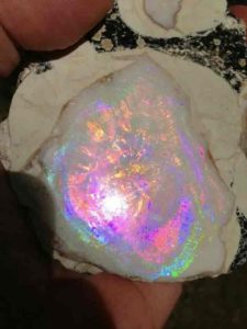
Geyser Opal “Spencer Opal” was formed in volcanic rocks around 4 million years ago by geysers. When a geyser would erupt, the water containing tiny silica spheres would create a thin layer of precious opal.
The main source of Spencer Opal Mines is the rhyolite and obsidian stream of gas pockets. The secondary deposit is a mixture of silica and water, collected inside the rock and released by a series of geysers. As a result of several eruptions over a period of time, the opal is deposited in layers. Most of the layers are thin, resulting in some of the most exquisite triplet opals in the world. Occasionally, the coating will be thin enough to slice a solid opal, which is an extra bonus for the finder.
How the Opal is formed
The main source of the mine is the rhyolite and obsidian stream of gas pockets. Opal Solution, or silica, was a secondary product for geyser activity. As a result of several eruptions over a period of time, the opal is deposited in layers. Most of the layers are thin, resulting in some of the most exquisite triplet opals in the world. Occasionally, the coating will be thin enough to slice a solid opal, which is an extra bonus for the finder.
Opal is hydrothermally deposited within hollow geodes in successive layers making Spencer Opal suitable for triplets as thin layers of extremely high quality precious opal are very translucent with a high color intensity. Opal doublets, often used in jewelry, are thin slices of precious opal attached to a matrix material. These gems are considerably cheaper than solid opals, but they provide the same color play. Opal doublets are sometimes coated with a thin layer or a transparent Quartz dome to make them more resistant to scratches (opal is a relatively soft gem). They’re known as triplets.
To make a triplet, the opal is ground flat parallel to the flame layer until the intense fire appears completely across the bottom. Then a flat piece of black Basinite or Obsidian is evaporated into a flat flame layer. The opal is again ground flat on the opposite side until the flame surface is revealed and reveals its maximum intensity against the black background. The opal should be about the thickness of the hair at this time. The Crystal cap is coated with epoxy and the opal is ground to its final form. This is how the Idaho Opal Triplet is made.
Colors of Opals and Their Vaule
Gems are taken from both the precious and the common forms, but the precious opal is the primary gem of this stone. There are many types of precious and common opals. The most attractive and beautiful type of opal is black opal, which is opal with a dark blue, dark green or black colored background. Next in value is white opal, which is opal with a light colored body color (black, green, cream, etc.) with a heavy color game. The next line is Mexican fire opal, a transparent to translucent orange red form of common opal. When Mexican fire opals exhibit colored game, they are known as Precious Fire Opal. In addition to being categorized as either black or white opals, most precious opals are further classified on the basis of the distribution and habit of their color play. There are also many names of varieties given to the numerous forms of both precious opal and common opal.
Where is spencer idaho?
Spencer is a city in Clark County, Idaho, United States. The population was 38 at the 2000 census. The city is noted as being the “Opal Capital of America”.[citation needed] The mine is located 5–6 miles outside of town.
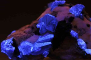
Benitoite crystals under UV light. Dallas Gem Mine (Benitoite Mine ; Benitoite Gem Mine ; Gem Mine), Dallas Gem Mine area, San Benito River headwaters area, New Idria District, Diablo Range, San Benito Co., California, USA. Photo Credit: Parent Géry
What is Benitoite Mineral?
Benitoite is a rare minerals best known to be the official Californian state gemstone. It is a barium titanium silicate mineral, typically of a blue color, found in hydrothermal rocks with a chemical composition of Ba Ti(Si 3O9). It is an extremely rare rock.
It is an extremely rare titanium cyclosilicate blue barium found in serpentinite that has been altered hydrothermally. This forms environments characteristic of subduction zones at convergent plate boundaries in low temperature, high pressure settings. Under short wave ultraviolet light, benitoite fluoresces which appear bright blue to bluish white in colour. Under long-wave UV light, the clearer to white benitoite crystals fluoresce green.
It was discovered about halfway between San Francisco and Los Angeles by prospector James M. Couch in the San Benito Mountains in 1907. Originally, Couch assumed that the mineral was a corundum mineral called sapphire because of its color similarity. A specimen was sent to the University of California, Berkeley in 1909 where mineralogist Dr. George D. Louderback discovered it was a mineral that was previously unknown. Corundum (sapphire) has a Mohs hardness specified at 9, while benitoite is much softer. He named it benitoite for its occurrence near the headwaters of the San Benito River in San Benito County, California.
It occurs globally in a variety of isolated locations, but only in California at the Benito Gem Mine where it was first discovered was gemstone quality material found. It has been correctly identified in Montana, Arkansas, Japan, and Australia even though they have evolved under slightly different conditions and are only increasing large enough to be considered a mineral accessory. Benitoite was named California’s official state gem in 1985.
It has a rare twinned crystal shape of 5 points, and an even rarer twinned crystal shape of 6 points, “David’s star,” with precisely 24 known specimens making it more precious than diamonds.
Physical Characteristics of Benitoite
Color: is typically blue, but also colorless and yellowish. Luster: is vitreous. Transparency: Crystals are transparent to translucent. Cleavage: is absent. Fracture: is irregular. Hardness: is 6 – 6.5 Specific Gravity: is approximately 3.6 (above average) Streak: is white
How is Benitoite formed?
It occurs only in the most mineralized region of California, the New Idria Mining District, in San Benito Country / USA (more than 150 semi-precious minerals and gemstones). There is a mountain range of basalt, chert, gray wack, shale, serpentine and Cretan and Tertiarian sandstones at the southern end or at the “Diablus Range.” It is a mountainous belt. Benitoite crystals have been formed from hydrothermal solutions containing relatively high levels of rare components such as barium, titanium, fluorine, iron etc. The minerals Albite, joaquinite, natrolite, neptunite and serpentine occur mainly with this. Natrolite, the last mineral produced at the deposit of benitoite, covered all of that deposit’s minerals. It is therefore necessary to remove the natrolite with acid (HCl) to expose the crystals of benitoite.
Where can benitoite be found?
Typically, benitoite occurs in combination with minerals which make up the host rock. The most widely used minerals are: natrolite, neptunite, joaquinite, serpentine and albite.
It is a rare mineral found in very few areas, including California, San Benito County, Japan and Arkansas. In the event of San Benito, it is contained in natural veins in a serpentinite body in glaucophane schists. The Mineral in Japan occurs on a dike cutting a serpentinite body of magnesio-riebeckite-quartz-phlogopite.
Where can I find Benitoite in California?
The Gem Benitoite mine is located about halfway between Los Angeles and San Francisco and has a small mining claim (Dallas claim) in San Benito County, California.
Why is benitoite a state gem in california?
Benitite was designated the official staple in 1985 by California.
It often referred to as the “blue diamond,” a very rare gem that varies in its hue, from light transparent to dark blue, to saphire blue or sometimes violet shad, which was found first on San Benito River headwater in 1907 (which is why the name is named).
It (pronounced as “benee-toe-ite”) was initially considered a saphire, but was found to be a science new mineral after careful analysis. Benitoite crystals of value are only found in California.
How much is Benitoite worth?
This gem is quite expensive Rich blue, clean stones at a carat and above sizes will command high prices. Clarity improves quality, particularly eye clean or better scores. Very light and very dark rocks lie at the lower end of the price continuum. At the top, you’ll find medium-dark stones.
Benitoite – $4,000 per carat
What is Benitoite used for?
Benitoite is used for a number of different purposes
The precious rock is used to produce jewellery as a gemstone.
Benitoite is another important use as a mineral sample. The mineral’s unique crystals are of great value as collectable samples.
This is also used as a small barium and titanium deposit.
How do you identify Benitoite?
This occurs in low temperature, high-pressure conditions typical of subduction zones at the edge of the converging plate. Under short wave ultraviolet light, benitoite fluoresces which appear bright blue to bluish white in colour. Under long-wave UV light, the clearer to white benitoite crystals fluoresce green.
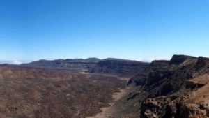
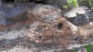
A detail of the southern wall of the Las Cañadas caldera de . It is the best exposed sector of this volcanic structure. (Imagen: Joan Martí)
Las Cañadas caldera (Tenerife, Canary Islands) is the result of different episodes of caldera collapses, associated to large explosive eruptions that triggered several landslides that modified the shape of the walls of the Las Cañadas volcanic edifice, according to a new review study published recently in the journal Earth-Sciences Reviews. The study integrates in the same solution the two main proposed hypothesis that have focused the long lasting scientific debate about the origin of this volcanic caldera where Teide-Pico Viejo active volcanic complex is located.
Joan Martí, researcher at the Institute of Earth Sciences Jaume Almera (Spanish National Research Council, ICTJA-CSIC), is the lead author of this new review article which aims to offer a “plausible explanation” for the origin of Las Cañadas caldera based on the current evidences gathered from all previous studies. “This volcanic structure is a chain of large scale destructive events that have accompanied the evolution of Tenerife Island”, summarizes Joan Martí.
The origin of the Las Cañadas caldera, named World Heritage Site by the UNESCO in 2007, has been controversial amongst the scientific community. Today it continues to be the focus of scientific debate. At present, there are two competing hypotheses.
The first idea suggests that the Las Cañadas caldera was a classic example of caldera collapse, when the magma chamber deep beneath the volcano drained abruptly during several explosive eruptions and the bedrock above collapsed over the emptied reservoir. According to this idea, its origin has occurred through one or several collapses of the volcanic edifice over the course of one or more explosive eruptions.
The second idea proposes that the caldera was the head of a large landslide. This idea suggests that a landslide occurred on a flank of Las Cañadas volcanic edifice due to gravitational instability, leading to the formation of the Icod valley.
The new article notes that “the apparent contradiction between these two hypotheses can be solved in a simple way: by admitting the existence of both processes which could have been temporally and even mechanically related”.
Joan Martí gathered and reviewed available information on the Las Cañadas caldera from 55 previous scientific studies. The ICTJA-CSIC researcher synthesised a large range of datasets, including stratigraphic, geochronological, structural, geomorphological, petrological, geophysical, borehole and bathymetric data.
Joan Martí explains that “in the case of Las Cañadas caldera, some of the clear geological clues and important data have been misinterpreted”. The ICTJA-CSIC researcher adds that “in this work, we have clarified the meaning of each of published evidences, locating them into the proper volcanological context”.
The article states that the amount and quality of data available geological and geophysical “should be sufficient to definitively close the controversy over the origin of this caldera”.
According to Joan Martí, this work offers “new starting points for future studies, because there are many things to study from this structure”. The researcher considers that “there is a lack of knowledge about the origin of the magmas that caused the explosive volcanism that led to the formation of the caldera, about the collapse mechanisms or even about the relationships between collapse calderas and large volume landslides”.
Deciphering the origin of this caldera “has important implications for risk assessment on Tenerife Island and also for the knowledge of the fresh water resources of the island, two aspects that can vary a lot depending on the adopted genetic model of the caldera”, concludes Joan Martí.
Reference:
Martí, J. (2019). Las Cañadas caldera, Tenerife, Canary Islands: A review, or the end of a long volcanological controversy. Earth-Science Reviews, 196, 102889. DOI:10.1016/j.earscirev.2019.102889
A panoramic view of one of the Arico ignimbrite outcrops where the two units can be seen.(Image: Joan Martí)
The emplacement of small-volume (<0,1km3) pyroclastic flows is strongly controlled by topography, according to a new study made by researchers of the Institute of Earth Sciences Jaume Almera of the Spanish National Research Council (ICTJA-CSIC) and University of Barcelona. The paper has been published in the journal Sedimentology.
The work has focused on the study and characterization of the Arico ignimbrite, located in the southern slopes of the Las Cañadas volcanic complex (Tenerife, Canary Islands). These rock formations were originated by the deposition of a 670.000 years old pyroclastic flow.
Guajara was the emission zone of this cloud made up of a mix of hot gases, volcanic ashes and rock fragments. This cloud showed fast downslope movement, and finally was deposited in the valleys of the southern zone of the island forming the studied ignimbrites.
“It is well known that pyroclastics flows are density currents controlled by the gravity and thus they tend to flow through valleys or depressed topographical zones”, explains Joan Martí, researcher at ICTJA-CSIC and first author of the study. “Now we have been able to demonstrate that the emplacement of small-volume pyroclastic flows is controlled by, besides the slope, the shape of the channel through which they flow. The bedrock morphology, obstacles, sudden slope variations or changes in the channel width are some of the topographical drivers that influence the emplacement and deposition of these types of pyroclastic flows”.
To do this, the team conducted fieldwork in the Barranco de los Ovejeros where they found and described a total of 57 outcrops. In this valley, the ignimbrite deposits are well exposed. Researchers studied the lithology, stratigraphy and the sedimentological features of the ignimbrite outcrops. They also measured the slope of the previous ground, depth and width of the channels that guided the transport and the final emplacement of the flow. Researchers also took 41 samples to conduct a paleomagnetic study to estimate the emplacement temperature of the Arico ignimbrite.
According to Joan Martí, despite its age, the Arico ignimbrites is a “well preserved and exposed deposit that permitted to reconstruct the paleo-topography with precision. Therefore, we were able to observe how the lithological, stratigraphical and sedimentary features of the ignimbrite deposits vary on the basis of the changes of the valley’s shape where they were finally emplaced.”
With all the field data, researchers were able to develop a theoretical model that explains the critical mechanisms from which topography guided the emplacement of the flow.
“Besides the usual parameters that define the flow regime of this type of volcanic material current, our model includes the contour conditions imposed by a particular topography which allows establishing how the flow conditions vary along with its emplacement”, notes Joan Martí.
According to the authors, this new model is “of general applicability and will help to explain other deposits of similar characteristics”.
Researchers describe in their study the different features of Arico ignimbrites along 7 observation points.
The paper notes that in the areas closest to the emission centre, the pyroclastic flow was efficiently channelled by the existing valleys that acted as efficient conduits. In these areas, the ignimbrite corresponds to a homogeneous moderately welded deposit.
The study states also that in the intermediate zones significant changes occurred in the steepness of the slope and here, although still channelled, the flow was influenced by hydraulic jumps, which controlled the way it was emplaced. In this area, two different sedimentary units can be clearly seen in the ignimbrite: the lower unit is orange, and the upper unit is grey.
Finally, in the distal areas near the present-day coastline, where the slope is very gentle or null and without a channelling relief that provoked a radial dispersion and fast degassing of the flow, only the upper unit can be found.
The researchers said that this work allows to a better understanding of the emplacement mechanisms of ignimbrites and to improve the volcanic hazard assessment. “This study opens the door to forecast the flow regime of the small-volume pyroclastic flows as long as we know the previous topography”.
Dario Pedrazzi and Domenico Doronzo, both researchers at ICTJA-CSIC, and Ferran Colombo, from the Faculty of Earth Sciences of the University of Barcelona, are the other authors of this new study.
Reference:
Martí, J. , Doronzo, D. M., Pedrazzi, D. and Colombo, F. (2019), Topographical controls on small‐volume pyroclastic flows. Sedimentology. DOI:10.1111/sed.12600
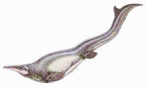
Plotosaurus bennisoni is a mosasaur from the Upper Cretaceous (Maastrichtian) North America. Restoration illustration from Wikimedia Commons, CC BY 3.0.
Mosasaurs were true sea monsters of late Cretaceous seas. These marine lizards — related to modern snakes and monitor lizards — grew as long as fifty feet, flashed two rows of sharp teeth, and shredded their victims with enormous, powerful jaws.
Now, new research suggests that mosasaurs had yet another potent advantage: a muscular breast stroke that may have added ambush-worthy bursts of speed.
“We know that mosasaurs most likely used their tails for locomotion. Now we think that they also used their forelimbs, or their tail and forelimbs together,” explains lead author Kiersten Formoso, a Ph.D. student in vertebrate paleontology at the University of Southern California. That dual swimming style, she says, could make mosasaurs unique among tetrapods (four limbed creatures), living or extinct.
Previous studies noted that mosasaurs had an unusually large pectoral girdle — the suite of bones that support the forelimbs. But most assumed the creature’s swimming was mainly driven by their long tails, something like alligators or whales. That smooth, long distance-adapted swimming style is called “cruising,” as opposed to “burst” motion. “Like anything that swims or flies, the laws of fluid dynamics mean that burst versus cruising is a tradeoff,” explains co-author Mike Habib, Assistant Professor of Anatomical Sciences at USC. “Not many animals are good at both.”
To dive in more closely on whether mosasaurs were burst-adapted, cruise-adapted, or an unusual balance of both, Formoso and co-authors focused on the oversized pectoral girdle. They studied a fossil Plotosaurus, a type of mosasaur, at the Natural History Museum of Los Angeles County. In addition, they used measurements of mosasaur pectoral girdles published in other studies.
They determined that the mosasaurs’ unusually large and low-placed pectoral girdle supported large muscle attachments. In addition, says Habib, asymmetry in the bone structure is a telltale sign of the strong, inward pull-down motion called adduction. These analyses suggest that mosasaurs used their forelimbs to swim, breast-stroke style, adding powerful bursts of propulsion to their ability to cruise.
The team continues to model bone structure, morphology, measurements, and fluid dynamics such as drag to learn exactly how, and how fast, these sea monsters swam. Along with applications to biomechanics, and even robotics, say Formoso and Habib, the study also sheds light on how evolution and ecosystems are affected by fluid dynamics.
Formoso points out that it’s a challenge to study kinematics on extinct animals, considering that the subjects are missing flesh, skin, and many bones. But one thing is nearly certain, she says. “Mosasaurs swam unlike anything else.”
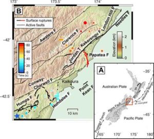
Fig. 1 Map of the 2016 Kaikōura earthquake and surrounding area. (A) Transpressional tectonic setting of the northeastern South Island of New Zealand. (B) Map of surface ruptures from the 2016 Mw 7.8 Kaikōura earthquake, shown in bold black lines with the Papatea fault in red (8, 28). Dots represent scaled relative energy release from back-projection results (15) and are colored by time since rupture onset. Mapped active faults that did not rupture during the Kaikōura event are indicated by thin black lines (28). Credit: Science Advances 02 Oct 2019: Vol. 5, no. 10, eaax5703, DOI: 10.1126/sciadv.aax5703
One of the 24-plus faults that ruptured in the 2016 magnitude 7.8 Kaikōura earthquake has turned out to be even more unusual than scientists first thought and it may prompt a rethink about how seismic hazard is calculated.
The Papatea Fault, unmapped before the quake and running along a similar path to the lower Clarence River in Marlborough, produced a 19km-long surface rupture and shunted a large area of mountainous country up by 8m in a matter of seconds.
A study published this week in the journal Science Advances indicates that the fault ruptured even though it hadn’t accumulated stress normally associated with fault rupturing.
Co-author and earthquake geologist at GNS Science Rob Langridge says it appears the fault suddenly became squeezed for room by the rupture of neighbouring faults causing it to break in “a very emphatic way.”
“The rupture of the Papatea Fault stands out for being one of the most dramatic elements of what was an unusual rupture sequence in the first place,” Dr. Langridge said.
“It produced the largest vertical movements of all the faults that ruptured during the earthquake and it has puzzled scientists because its rupture could not be fitted to standard models of fault rupture.”
However, the paper published this week used computer analysis of LIDAR images to come up with a solution to its unusual behavior. It was written by Canadian MSc student Anna Diederichs with colleague and geophysicist Ed Nissen, both of the University of Victoria in British Columbia. Three scientists from GNS Science, including Langridge, were co-authors.
“We discovered a number of unusual characteristics to this fault. Most unusually, the standard elastic rebound model of earthquake faulting did not fit the observed ground deformation,” Dr. Nissen said.
“We’ve concluded that the Papatea Fault did not release elastically stored tectonic strain as faults normally do during a rupture.”
Dr. Nissen said the findings indicate that some faults may fall outside typical fault behavior and conventional modeling may not capture the hazard they pose.
He said earthquake forecasting is based on the elastic strain cycle model where faults gradually accumulate strain until they fail, and then the cycle is repeated.
“However, the Papatea Fault does not seem to follow this model, and such faults may still need to be accounted for in earthquake forecast models.”
Going forward, he said this research finding could be considered when assessing the risk from faults that might have a low or unclear strain accumulation signal.
The research was based on computer analysis of pre- and post-earthquake LIDAR images of the fault rupture area. Fortuitously, Environment Canterbury collected LIDAR of the Clarence Valley area several years before the Kaikōura earthquake mainly for flood protection purposes. These images were compared with LIDAR images collected in the wake of the earthquake in 2016.
Reference:
A. Diederichs et al. Unusual kinematics of the Papatea fault (2016 Kaikōura earthquake) suggest anelastic rupture, Science Advances (2019). DOI: 10.1126/sciadv.aax5703
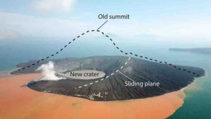
A drone image two weeks after the flank collapsed. The volcano peak that had measured 320 meters is missing. Credit: GFZ
On 22 December 2018, a flank of the Anak Krakatau volcano plunged into the Sunda strait between the Indonesian islands of Sumatra and Java, triggering a tsunami that killed 430 people. An international research team led by Thomas Walter of the German Research Centre for Geosciences GFZ in Potsdam has now shown that the volcano produced clear warning signals before its collapse. This was the result of the analysis of a large amount of data from very different sources collected during ground-based measurements as well as by drones and satellites. Satellite data, for example, showed increased temperatures and ground movement on the southwestern flank months before the catastrophe. Seismic data and low-frequency sound waves from a smaller earthquake two minutes before the sudden collapse of a large part of the volcano had heralded the fatal event. This collapse finally triggered the deadly tsunami. The researchers want to use the analysis of this complex event cascade to improve monitoring and early detection of other volcanoes. Their study was published in the journal Nature Communications.
Volcanic islands like Anak Krakatau often consist of unstable material. Therefore, every now and then a collapse of volcanic flanks occurs on these islands. Yet, this had not been precisely measured until now. “At Krakatau, we were able to observe for the first time how the erosion of such a volcanic flank took place and which signals announced it,” Thomas Walter, a volcanologist at the GFZ explains. In their study at Anak Krakatau the researchers were able to show that over months the movement of the southeast flank towards the sea formed a kind of slide. The sudden accelerated slide of the flank into the sea, the so-called flank collapse, lasted only two minutes and was measured by seismographs and infrasound networks before the first impacts of the tsunami had reached the coasts.
“We used an exceptionally broad range of methods: From satellite observation to ground-based seismic data, from infrasound to drone data, from temperature measurements to chemical analysis of eruption products,” says Thomas Walter. “Today’s almost unrestricted access to worldwide data was critical in this. In the days following the tsunami, it allowed us to analyse this event at different locations in different countries at the same time.”
Improved monitoring systems as a goal
Similar to Anak Krakatau such events could also herald themselves on other volcanic islands in the Atlantic, Pacific or even in the Mediterranean, to which the results of the study could then presumably be transferred, according to Walter. “We assume that tsunami early warning systems must also take into account events caused by landslides. Those volcanoes that are at risk of slipping should be integrated into the monitoring systems.”
Seismologist Frederik Tilmann from GFZ and Freie Universität Berlin was also involved in the study. He says that the unusual seismic pattern of the flank collapse was a particular challenge when analysing the data. In contrast to tectonic earthquakes, only a small part of this pattern consisted of high frequencies around 1 Hertz (1 oscillation per second). Instead, the earthquake waves contained stronger components in the range of low frequencies up to about 0.03 Hertz (1 oscillation per 35 seconds). “This property was the reason why the event was not detected in any routine evaluation,” says Tilmann.
The effort of monitoring systems will pay back, since a large part of the victims of volcanoes in the past two centuries have not been killed by the eruptions themselves, but by landslides and tsunamis, according to Walter. The new results show that the danger of collapsing volcanoes has so far been underestimated. The first step now is to identify the volcanoes at particular risk and to supplement existing measurement methods with additional sensors and new algorithms for evaluation. “We are confident that our findings will lead to the development of improved monitoring systems,” said Walter.
Reference:
Thomas R. Walter, Mahmud Haghshenas Haghighi, Felix M. Schneider, Diego Coppola, Mahdi Motagh, Joachim Saul, Andrey Babeyko, Torsten Dahm, Valentin R. Troll, Frederik Tilmann, Sebastian Heimann, Sébastien Valade, Rahmat Triyono, Rokhis Khomarudin, Nugraha Kartadinata, Marco Laiolo, Francesco Massimetti, Peter Gaebler. Complex hazard cascade culminating in the Anak Krakatau sector collapse. Nature Communications, 2019; 10 (1) DOI: 10.1038/s41467-019-12284-5
The discovery of a previously unknown species of pterosaur, which may have persisted as late as the Turonian period (90-93 million years ago), is reported in Scientific Reports this week. The fossil, which includes parts of the skull and five vertebrae, is the most complete pterosaur specimen ever found in Australia. The findings suggest it may be a late-surviving member of the Anhanguera genus of pterodactyls, which were believed to have gone extinct at the end of the Cenomanian period (100-94 million years ago).
Pterosaurs are known from fossils discovered on every continent but their remains are often incomplete and fragmentary because their bones are thin and hollow. The fossil record for pterosaurs in Australia is particularly sparse with only 20 known fragmentary specimens.
Adele Pentland and colleagues discovered the new pterosaur, which they have named Ferrodraco lentoni (from the Latin ferrum (iron), in reference to the ironstone preservation of the specimen, and the Latin draco (dragon)), in the Winton Formation of Queensland. Based on the shape and characteristics of its jaws, including crests on upper and lower jaw and spike-shaped teeth, the authors identified the specimen as belonging to the Anhanguera, which are known from the Early Cretaceous Romualdo Formation of Brazil. Comparison with other anhanguerian pterosaurs suggests that Ferrodraco’s wingspan measured approximately four metres. The authors also report a number of unique dental characteristics, including small front teeth, which distinguish Ferrodraco from other anhanguerians and identify it as a new species.
The fossil was discovered in 2017 in a part of the Winton formation that may have formed as late as the early Turonian, which suggests that the anhanguerians may have survived later in Australia than elsewhere.
Reference:
Ferrodraco lentoni gen. et sp. nov., a new ornithocheirid pterosaur from the Winton formation (cenomanian-lower turonian) of Queensland, Australia, DOI: 10.1038/s41598-019-49789-4
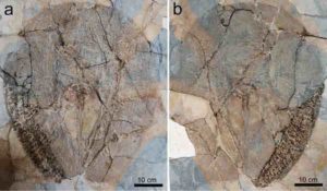
One of the three fossils of Lessiniabatis aenigmatica (MNHN F.Bol.566) from the famous fossil site of Monte Bolca (Italy) preserved as part and counterpart. The specimen is housed in the Museum National d’Histoire Naturelle of Paris. Credit: Giuseppe Marramà
An international research team led by Giuseppe Marramà from the Institute of Paleontology at the University of Vienna discovered a new and well-preserved fossil stingray with an exceptional anatomy, which greatly differs from living species. The find provides new insights into the evolution of these animals and sheds light on the recovery of marine ecosystems after the mass extinction occurred 66 million years ago. The study was recently published in the journal Scientific Reports.
Stingrays (Myliobatiformes) are a very diverse group of cartilaginous fishes which are known for their venomous and serrated tail stings, which they use against other predatory fish, and occasionally against humans. These rays have a rounded or wing-like pectoral disc and a long, whip-like tail that carries one or more serrated and venomous stings. The stingrays include the biggest rays of the world like the gigantic manta rays, which can reach a “wingspan” of up to seven meters and a weight of about three tons.
Fossil remains of stingrays are very common, especially their isolated teeth. Complete skeletons, however, exist only from a few extinct species coming from particular fossiliferous sites. Among these, Monte Bolca, in northeastern Italy, is one of the best known. So far, more than 230 species of fishes have been discovered that document a tropical marine coastal environment associated with coral reefs which dates back to about 50 million years ago in the period called Eocene.
This new fossil stingray has a flattened body and a pectoral disc ovoid in shape. What is striking is the absence of sting and the extremely short tail, which is not long as in the other stingrays, and does not protrude posteriorly to the disc. This body plan is not known in any other fossil or living stingray. Since this animal is unique and peculiar, the researchers named the new stingray Lessiniabatis aenigmatica, which means “bizarre ray from Lessinia” (the Italian area where Bolca is located).
More than 70 percent of the organisms, such as dinosaurs, marine reptiles, several mammal groups, numerous birds, fish and invertebrates, disappeared during the fifth largest extinction event in the Earth’s history occurred about 66 million years ago at the end of the Cretaceous. In marine environments, the time after this event is characterized by the emergence and diversification of new species and entire groups of bony and cartilaginous fishes (sharks and rays), which reoccupied the ecological niches left vacant by the extinction’s victims. The new species experimented sometimes new body plans and new ecological strategies.
“From this perspective, the emergence of a new body plan in a 50-million-year-old stingray such as Lessiniabatis aenigmatica is particularly intriguing when viewed in the context of simultaneous, extensive diversification and emergence of new anatomical features within several fish groups, during the recovery of the life after the end-Cretaceous extinction event,” states Giuseppe Marramà.
Reference:
Giuseppe Marramà et al, A bizarre Eocene dasyatoid batomorph (Elasmobranchii, Myliobatiformes) from the Bolca Lagerstätte (Italy) reveals a new, extinct body plan for stingrays, Scientific Reports (2019). DOI: 10.1038/s41598-019-50544-y
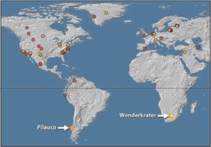
A world map that shows where similary platinum spikes have been discovered in the world. The latest discovery is at the Wonderkrater site in Limpopo Province, South Africa. Credit: Francis Thackeray/Wits University
A team of scientists from South Africa has discovered evidence partially supporting a hypothesis that Earth was struck by a meteorite or asteroid 12 800 years ago, leading to global consequences including climate change, and contributing to the extinction of many species of large animals at the time of an episode called the Younger Dryas.
The team, led by Professor Francis Thackeray of the Evolutionary Studies Institute at the University of the Witwatersrand in Johannesburg, South Africa, discovered evidence of a remarkable “platinum spike” at a site called Wonderkrater in the Limpopo Province, north of Pretoria in South Africa. Working with researcher Philip Pieterse from the University of Johannesburg and Professor Louis Scott of the University of the Free State, Thackeray discovered this evidence from a core drilled in a peat deposit, notably in a sample about 12 800 years old. This research was published in Palaeontologia Africana.
Noting that meteorites are rich in platinum, Thackeray said “Our finding at least partially supports the highly controversial Younger Dryas Impact Hypothesis (YDIH). We seriously need to explore the view that an asteroid impact somewhere on earth may have caused climate change on a global scale, and contributed to some extent to the process of extinctions of large animals at the end of the Pleistocene, after the last ice age.”
Many mammals became extinct in North America, South America and Europe at the time of the Younger Dryas. In South Africa a few extraordinary large animal species became extinct, not necessarily at exactly 12 800 years ago, but close to that period. These megafauna include a giant African buffalo, a large zebra, and a very big wildebeest.
Human populations may also have been indirectly affected at the time in question. In North America there is a dramatic termination of the stone tool technology of Clovis people. Remarkably, archaeologists in South Africa have detected an almost simultaneous termination of the Robberg stone artifact industry associated with people in some parts of the country, including the area around Boomplaas near the Cango Caves in the southern Cape, close to the town of Oudshoorn.
“Without necessarily arguing for a single causal factor on a global scale, we cautiously hint at the possibility that these technological changes, in North America and on the African subcontinent at about the same time, might have been associated indirectly with an asteroid impact with major global consequences,” says Thackeray. “We cannot be certain, but a cosmic impact could have affected humans as a result of local changes in environment and the availability of food resources, associated with sudden climate change.”
At Wonderkrater, the team has evidence from pollen to show that about 12 800 years ago there was temporary cooling, associated with the “Younger Dryas” drop in temperature that is well documented in the northern hemisphere, and now also in South Africa. According to some scientists, this cooling in widespread areas could at least potentially have been associated with the global dispersal of platinum-rich atmospheric dust.
A large crater 31 kilometers in diameter has been discovered in northern Greenland beneath the Hiawatha Glacier. “There is some evidence to support the view that it might possibly have been the very place where a large meteorite struck the planet earth 12 800 years ago,” says Thackeray. “If this was indeed the case, there must have been global consequences.”
Thackeray’s team believes their discovery of a platinum spike at about 12 800 years ago at Wonderkrater is just part of the strengthening view that an asteroid or cometary impact might have occurred at that time.
This is the first evidence in Africa for a platinum spike preceding climate change. Younger Dryas spikes in platinum have also been found in Greenland, Eurasia, North America, Mexico and recently also at Pilauco in Chile. Wonderkrater is the 30th site in the world for such evidence.
“Our evidence is entirely consistent with the Younger Dryas Impact Hypothesis” says Thackeray.
The discovery in South Africa is expected to be integrated with those made in other parts of the world, recognizing that the source of the platinum at Wonderkrater could hypothetically be cosmic dust that was dispersed in the atmosphere after a meteorite impact in Greenland.
The probability of a large asteroid striking Earth in the future may seem to be low, but there are thousands of large rocks distributed primarily between Jupiter and Mars. One in particular, classified as Apophis 99942, is referred to as a “Potentially Hazardous Asteroid.” It is 340 meters wide and will come exceptionally close to the Earth in 10 years’ time.
“The closest encounter will take place precisely on Friday April 13, 2029,” says Thackeray. “The probability of the Apophis 99942 asteroid hitting us then is only one in 100 000, but the probability of an impact may be even higher at some time in the future, as it comes close to Earth every 10 years.”
Reference:
The Younger Dryas interval at Wonderkrater (South Africa) in the context of a platinum anomaly hdl.handle.net/10539/28129
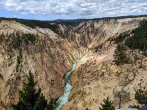
A river flows between mountains at Yellowstone National Park in Wyoming. New research tests a 30-year-old theory on the relationship between erosion and mountain height. Photo credit: Seth Cottle/Unsplash
Over millions of years, Earth’s summits and valleys have moved and shifted, resulting in the dramatic landscapes of peaks and shadows we know today. Mountains often form when pressure under Earth’s surface pushes upward, yet many factors impact their ultimate height, including the erosion of the areas between mountains, known as channels.
Scientists have long assumed that as land is pushed faster upward to form a mountain, its height increases in a continuous and predictable way. But new research shows that these predictions may stop working for the steepest mountains and therefore limit their height — and this may hold true for ranges on the entire planet.
“People have argued for a long time that as channels get steeper and steeper, the erosion rate keeps increasing,” said George Hilley, a professor of geological sciences at Stanford University’s School of Earth, Energy & Environmental Sciences (Stanford Earth) and lead author of a study published in Nature Geoscience Sept. 16. “We find that the theory works really well until a certain point and then it breaks down empirically — it seems as though something else kicks in that we don’t completely understand.”
The researchers analyzed samples from a broad range of mountain landscapes across the tropics, including Venezuela, Brazil, Guatemala, Costa Rica and Taiwan, controlling for rock type and climate conditions to assess parallel comparisons. They found that after mountains reach a certain elevation, channels between mountains suddenly become extremely sensitive to subtle changes in their inclines, thereby limiting the height of the mountains above. They added data from hundreds of mountain ranges worldwide and found they followed a similar pattern: the height, or relief, of the landscape is capped after crossing a threshold driven by channel steepness.
“Areas of land above channels are likely being controlled by how quickly a river can cut down — this is the framework by which we understand how the height of mountains varies as a function of climate and the collision of continents,” Hilley said. “The anomaly we observed is kind of a mystery and is not necessarily what conventional theory might predict.”
Research impacts
By bringing evidence of this mysterious factor influencing mountain height, the research could impact other work, such as studies on the relationship between mountain erosion rates and climate — important elements for understanding ancient climate and forecasting future patterns.
“Our work adds an interesting depth to some of these studies, because the way in which Earth’s topography changes as climates become more or less erosive may also change as the threshold is approached,” Hilley said.
The work also has implications for the geophysical links of mountain formation, which scientists are interested in exploring to understand the hidden activity of tectonic movements below our feet.
“People like me have always hoped that you could actually use the topography in order to say something about how quickly faults might be slipping,” Hilley said. “What our results say is that you can still do that in landscapes that are moderately steep, but it might become increasingly difficult as landscapes become steeper.”
Global patterns
The scientists took a fundamentally different approach to the research by searching the globe for conditions that reveal changes in mountain height rather than focusing on just one location. That search led them to focus on ranges in the tropics, but the conclusions were consistent across all regions of the globe and may also be applied to understand ancient topography.
“The Himalayas are being uplifted pretty rapidly and they expose pretty hard rocks, and indeed, when you measure them out, they are pretty close to this threshold,” he said. “You might be able to take this threshold with just the modern configuration of the landscape and actually place some upper bound on what the topography of the ancient Himalayas looked like.”
Hilley said the results of the study were surprising, as well as the fact that they remained consistent when compared with global data.
“In retrospect it makes sense when you look at it from the overall context of what our planet actually looks like,” Hilley said. “It really speaks to the fact that there might be lots of fertile ground to explore why this might happen. It also points to the fact that there might be something about the way in which rivers incise that we just don’t understand yet.”
Reference:
George E. Hilley, Stephen Porder, Felipe Aron, Curtis W. Baden, Samuel A. Johnstone, Frances Liu, Robert Sare, Aaron Steelquist, Holly H. Young. Earth’s topographic relief potentially limited by an upper bound on channel steepness. Nature Geoscience, 2019; DOI: 10.1038/s41561-019-0442-3
Subduction forced the younger oceanic crust down beneath the supercontinent Pangaea millions of years ago. Credit: University of Melbourne
Our planet formed around 4.54 billion years ago but few hints of this ancient world remain—just a small outcrop of rocks in northwestern Canada dating back 4.03 billion years and tiny crystals of the mineral zircon from western Australia that are about 4.3 billion years old.
The vast majority of the thin crust that we live on is considerably younger; this lack of preserved older material is a consequence of our dynamic planet.
The constant jostling of tectonic plates forms and destroys rocks, while the action of the hydrological cycle—rain, rivers, glaciers, oceans—tends to erode and redistribute their constituents.
For many decades, however, scientists have hypothesized that there are areas deep in the Earth’s interior that contain material untouched since the planet was formed.
These domains of primordial material are leftovers from the ancient event that saw the separation of our planet’s core from the silicate component which makes up most of the Earth’s crust and mantle.
Now, new University of Melbourne research is shedding some light on this puzzle using kimberlites—an igneous rock.
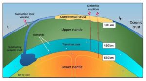
These unusual magmas are the primary source of one of our most treasured commodities—diamonds. They are the only volcanic deposits we know to have come from Earth’s deep mantle and they provide a fascinating glimpse into our planet’s formation.
Despite our best efforts, hypotheses about what lies deep in the Earth’s interior have remained largely untested.
We can create images of our planet’s interior using geophysical techniques involving seismic wave transmission, but it is a much harder task to determine the composition of the deep Earth.
Samples are rarely presented to us for analysis, and we do not have the technology to drill into the Earth’s mantle to find this material at its source.
The deepest hole ever drilled, the Kola Superdeep Borehole in northwest Russia, reaches just over 12 kilometers in depth.
Although that may sound deep, it’s barely one third of the way through the crust in that region. In fact, we would need to dig more than 500 kilometers further into the underlying mantle to have any chance of finding primordial material.
Many of our ideas about the composition of the Earth’s interior actually come from meteorites.
We believe they derive from catastrophic collisions that released material from deep within early solar system planets that were formed in a similar way to the Earth.
However, there are rare eruptive events that bring to the surface material from deep in the Earth, such as kimberlites.
Kimberlite eruptions have never been witnessed, because most kimberlites were formed millions to billions of years ago.
But we know from their textures and volatile-rich nature that these eruptions must have been extremely violent, traveling through the Earth’s mantle at extreme speed and sampling their surroundings as they went.
A small percentage of diamonds carry tiny inclusions of other minerals which are only stable at great pressures, providing clear evidence of their formation happening at depths of up to 800 kilometers.
In our study with University of Melbourne researchers Professor David Phillips and Drs Andrea Giuliani and Roland Maas, Professor Graham Pearson from the University of Alberta, and Dr. Geoff Nowell from Durham University, we measured the composition of kimberlites that erupted over a 2.5 billion year period of Earth history; collecting data and samples from thirteen kimberlite fields globally, spanning every continent except Antarctica.
Using sensitive radioisotope tracers, we can map the evolution of their mantle sources through time.
Our results show that, prior to around 200 million years ago, all kimberlites that erupted on Earth were likely sourced from a single primordial reservoir, formed soon after Earth’s core formed.
Then, around 200 million years ago, this reservoir appears to have been disrupted.
This was possibly due to a vast subduction zone established along the margins of the supercontinent Pangaea—the single continent that predated the seven continents that we now have.
Here, collision between tectonic plates forced the younger oceanic crust down beneath the supercontinent and back into the Earth’s deeper mantle. This material may have resulted in the contamination of the primordial reservoir.
These observations provide our best evidence yet for the existence of an early primordial reservoir within the Earth’s mantle—a subject of intense speculation for the last four decades.
And this major event around 200 million years ago may well have represented a significant turning point in the Earth’s geochemical evolution.
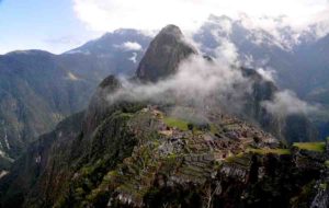
Machu Pichu. Detailed mapping indicates the World Heritage Site’s location and layout were dictated by the underlying geological faults. Photo taken 5 Nov. 2010; credit: Rualdo Menegat.
The ancient Incan sanctuary of Machu Picchu is considered one of humanity’s greatest architectural achievements. Built in a remote Andean setting atop a narrow ridge high above a precipitous river canyon, the site is renowned for its perfect integration with the spectacular landscape. But the sanctuary’s location has long puzzled scientists: Why did the Incas build their masterpiece in such an inaccessible place? Research suggests the answer may be related to the geological faults that lie beneath the site.
On Monday, 23 Sept. 2019, at the GSA Annual meeting in Phoenix, Rualdo Menegat, a geologist at Brazil’s Federal University of Rio Grande do Sul, will present the results of a detailed geoarchaeological analysis that suggests the Incas intentionally built Machu Picchu — as well as some of their cities — in locations where tectonic faults meet. “Machu Pichu’s location is not a coincidence,” says Menegat. “It would be impossible to build such a site in the high mountains if the substrate was not fractured.”
Using a combination of satellite imagery and field measurements, Menegat mapped a dense web of intersecting fractures and faults beneath the UNESCO World Heritage Site. His analysis indicates these features vary widely in scale, from tiny fractures visible in individual stones to major, 175-kilometer-long lineaments that control the orientation of some of the region’s river valleys.
Menegat found that these faults and fractures occur in several sets, some of which correspond to the major fault zones responsible for uplifting the Central Andes Mountains during the past eight million years. Because some of these faults are oriented northeast-southwest and others trend northwest-southeast, they collectively create an “X” shape where they intersect beneath Machu Picchu.
Menegat’s mapping suggests that the sanctuary’s urban sectors and the surrounding agricultural fields, as well as individual buildings and stairs, are all oriented along the trends of these major faults. “The layout clearly reflects the fracture matrix underlying the site,” says Menegat. Other ancient Incan cities, including Ollantaytambo, Pisac, and Cusco, are also located at the intersection of faults, says Menegat. “Each is precisely the expression of the main directions of the site’s geological faults.”
Menegat’s results indicate the underlying fault-and-fracture network is as integral to Machu Picchu’s construction as its legendary stonework. This mortar-free masonry features stones so perfectly fitted together that it’s impossible to slide a credit card between them. As master stoneworkers, the Incas took advantage of the abundant building materials in the fault zone, says Menegat. “The intense fracturing there predisposed the rocks to breaking along these same planes of weakness, which greatly reduced the energy needed to carve them.”
In addition to helping shape individual stones, the fault network at Machu Picchu likely offered the Incas other advantages, according to Menegat. Chief among these was a ready source of water. “The area’s tectonic faults channeled meltwater and rainwater straight to the site,” he says. Construction of the sanctuary in such a high perch also had the benefit of isolating the site from avalanches and landslides, all-too-common hazards in this alpine environment, Menegat explains.
The faults and fractures underlying Machu Picchu also helped drain the site during the intense rainstorms prevalent in the region. “About two-thirds of the effort to build the sanctuary involved constructing subsurface drainages,” says Menegat. “The preexisting fractures aided this process and help account for its remarkable preservation,” he says. “Machu Picchu clearly shows us that the Incan civilization was an empire of fractured rocks.”
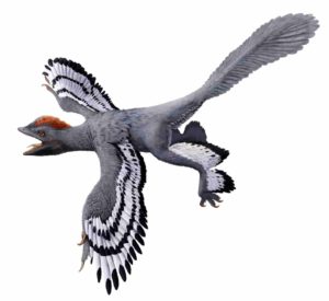
A life reconstruction of the feathered dinosaur Anchiornis huxleyi based on fossil evidence of its color and patterning. This evidence included inferences about melanin pigments. Credit: HKU MOOC / Julius T Csotonyi / Michael Pittman.
Dr Michael Pittman of the Vertebrate Palaeontology Laboratory, Department of Earth Sciences, The University of Hong Kong led an international study with his PhD student Mr Arindam Roy that evaluates fossil colour reconstruction methods to propose a new study framework that improves and expands current practice. The paper was recently published in the journal Biological Reviews.
“People are fascinated by the colour and pattern of dinosaurs and other extinct animals because these aspects can tell you so much about an animal. Just think of a zebra and a peacock. We evaluated everything we know about fossil and modern animal colour and used that knowledge to propose a framework to improve how we reconstruct fossil colour in the future.” said Dr Pittman.
Colour and patterns are critical to understanding the life, ecology, physiology and behaviour of animals. These colours are produced when light interacts with pigments and the structure of animal tissue. Common naturally-occurring animal pigments include melanin, carotenoids, porphyrins pterins, flavins and psittacofulvins which produce colours ranging from black and grey to yellow, orange and green.
Feathered dinosaur fossils instrumental to understanding the origin of birds were the first animal fossils to yield evidence of melanin, the colour pigment we also have in our eyes and hair. In the last ten years, colour patterns have been reconstructed in over 30 fossil animals including birds, non-avialan dinosaurs and mammals, providing a unique opportunity to test ecological and behavioral hypotheses that were previously out of reach. Unfortunately, our knowledge of other pigments is scarce in the fossil record as these non-melanin pigments are more difficult to fossilise. This incomplete knowledge and the lack of a standard study approach have been prevailing challenges to the reconstruction of colour in fossil animals.
Co-author Dr Evan Saitta of the Field Museum of Natural History, Chicago, USA said, “We are in the golden age of multidisciplinary techniques in palaeontology. This is the first comprehensive study that not only critically evaluates the currently available methods, but also provides a reliable and repeatable framework that covers all vertebrate pigment systems not just melanin alone.”
The new palaeocolour reconstruction framework proposed by Dr Michael Pittman, Mr Arindam Roy and their international team comprises four main steps: (1) Map the known or suspected extent of preserved colour and patterns in the specimen; (2) Search for pigment-bearing microstructures using electron microscopy e.g. microstructure shape can be used to identify melanin-based colours like black, grey and brown); (3) If melanin-based colours are not detected, use high-end chemical analysis techniques to detect biomarkers of other pigments (4) Use reconstructed colours and patterns to test fundamental hypotheses related to animal physiology, ecology and behaviour. The new framework overcomes past challenges by incorporating the chemical signatures of different pigments, large and small-scale anatomical details visible in fossils as well as the potential for different pigments to fossilise. This framework provides background context for the evolution of colour-producing mechanisms and is expected to encourage future efforts to reconstruct colour in more fossil animals including non-dinosaur reptiles and mammals.
Mr Roy, the study’s first author and a Hong Kong PhD Fellow said, “I am really excited by the course we have charted in this review study as I will be tackling many of the key issues we identified during my PhD studies at HKU.”
Reference:
Arindam Roy, Michael Pittman, Evan T. Saitta, Thomas G. Kaye, Xing Xu. Recent advances in amniote palaeocolour reconstruction and a framework for future research. Biological Reviews, 2019; DOI: 10.1111/brv.12552
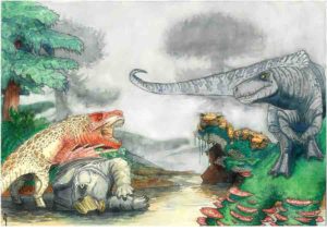
Artist’s reconstruction of two rauisuchians fighting over a desiccated corpse of a mammal-relative in the Triassic of southern Africa. In the background, dinosaurs and mammal-like reptiles form other parts of the ecosystem. Credit: Viktor Radermacher, owns copyright
“These ancient fossils provide us with evidence of how at least two predator species hunted these vegetarian dinosaurs 210 million-years-ago. It is amazing to follow the clues left behind in fossilised teeth, jaws, limbs and other fossils to help us tell the ancient story of life in southern Africa,” says Tolchard.
The fossils studied by Tolchard include teeth, pieces of jaws, hind limbs and body armour, all of which are can be described as parts of rauisuchians.
Rauisuchians are closely related to crocodiles as we know them today. They had a diversity of body shapes and sizes during the Triassic period. The specimens described in this research include some of the largest carnivorous members of this group, that were possibly up to 10 metres long, with huge skulls full of serrated, curved teeth.
The study, published online in the Journal of African Earth Sciences last week, shows that the rauisuchians were some of the latest-surviving members of their group, and that when they were alive, they were thriving close to the Antarctic Circle — the theoretical limit for their physiology.
“In the Triassic period, rauisuchians were widespread and their fossils are known from all continents except Antarctica,” adds Tolchard. “They went extinct about 200 million years ago, paving the way for dinosaurs to become the dominant large land animals.”
“Rick’s study demonstrates the value of re-examining old specimens, and now we finally know what was preying on all those herbivorous dinosaurs!” says Professor Jonah Choiniere, Rick’s advisor and Professor of Comparative Palaeobiology at the Wits Evolutionary Studies Institute.
Tolchard studied fossils from collections based at the the University of the Witwatersrand, the Iziko South African Museum and the National Museum in Bloemfontein. He was joined in the research by an international team, including researchers from the USA, Argentina and the UK.
Reference:
Frederick Tolchard, Sterling J. Nesbitt, Julia B. Desojo, Pia Viglietti, Richard J. Butler, Jonah N. Choiniere. ‘Rauisuchian’ material from the lower Elliot Formation of South Africa and Lesotho: Implications for Late Triassic biogeography and biostratigraphy. Journal of African Earth Sciences, 2019; 160: 103610 DOI: 10.1016/j.jafrearsci.2019.103610
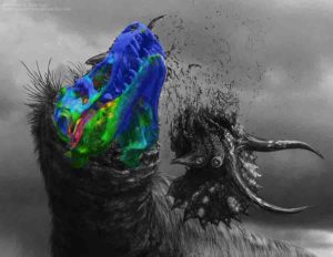
An artist’s rendition of the Tyrannosaurus rex with the 3D imaging showing muscle activation in its head. Illustration courtesy of Brian Engh.
A Tyrannosaurus rex could bite hard enough to shatter the bones of its prey. But how it accomplished this feat without breaking its own skull bones has baffled paleontologists. That’s why scientists at the University of Missouri are arguing that the T. rex’s skull was stiff much like the skulls of hyenas and crocodiles, and not flexible like snakes and birds as paleontologists previously thought.
“The T. rex had a skull that’s 6 feet long, 5 feet wide and 4 feet high, and bites with the force of about 6 tons,” said Kaleb Sellers, a graduate student in the MU School of Medicine. “Previous researchers looked at this from a bone-only perspective without taking into account all of the connections — ligaments and cartilage — that really mediate the interactions between the bones.”
Using a combination of imaging, anatomy and engineering analysis, the team observed how the roof of the mouth of the T. rex reacted to the stresses and strains from chewing by applying models of how two present day relatives of T. rex — a gecko and a parrot — chew to how the T. rex skull worked.
“Dinosaurs are like modern-day birds, crocodiles and lizards in that they inherited particular joints in their skulls from fish — ball and socket joints, much like people’s hip joints — that seem to lend themselves, but not always, to movement like in snakes,” said Casey Holliday, an associate professor of anatomy in the MU School of Medicine. “When you put a lot of force on things, there’s a tradeoff between movement and stability. Birds and lizards have more movement but less stability. When we applied their individual movements to the T. rex skull, we saw it did not like being wiggled in ways that the lizard and bird skulls do, which suggests more stiffness.”
In addition to helping paleontologists with a detailed study of the anatomy of fossilized animals, researchers believe their findings can help advance human and animal medicine by providing better models of how joints and ligaments interact.
“In humans, this can also be applied to how people’s jaws work, such as studying how the jaw joint is loaded by stresses and strains during chewing,” said Ian Cost, the lead researcher on the study. Cost is an assistant professor at Albright College and a former doctoral student in the MU School of Medicine. “In animals, understanding how those movements occur and joints are loaded will, for instance, help veterinarians better understand how to treat exotic animals such as parrots, which suffer from arthritis in their faces.”
Reference:
Ian N. Cost, Kevin M. Middleton, Kaleb C. Sellers, Michael Scott Echols, Lawrence M. Witmer, Julian L. Davis, Casey M. Holliday. Palatal Biomechanics and Its Significance for Cranial Kinesis in Tyrannosaurus rex. The Anatomical Record, 2019; DOI: 10.1002/ar.24219
Yellowstone National Park is an American national park located in Wyoming, Montana, and Idaho.
Places To Visit in United States of America
In this Article you will find some of the best natural Places To Visit in United States of America
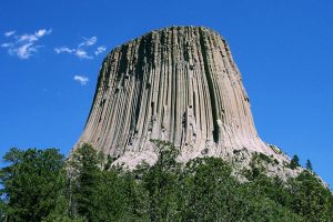
1. Devils Tower, Wyoming
Devils Tower. Photo Copyright: Wikipedia
Devils Tower (also recognized as Bear Lodge Butte) is a butt, potentially laccolithic, made up of igneous rock in the Black Hills Bear Lodge Ranger District, close Hulett and Sundance in Crook County, northeast of Wyoming, overlooking the Belle Fourche River. It rises above the Belle Fourche River at 1,267 feet (386 m), sitting at 867 feet (265 m) from top to bottom. The peak is 5,112 feet above sea level (1,559 m).
Devils Tower was the first National Monument to be founded by President Theodore Roosevelt on September 24, 1906. The border of the monument encompasses an area of 1,347 acres (545 ha).
Approximately 1% of the 400,000 annual tourists to the monument have visited the Devils Tower in latest years, mostly using traditional climbing methods and it is the best place to visit in united states of america.
The name Devil’s Tower arose during an expedition led by Colonel Richard Irving Dodge in 1875, when his translator allegedly misinterpreted a indigenous word meaning “Bad God’s Tower.” All data advertisements in that region use the word “Devils Tower,” following a norm of geographical naming that omits the apostrophe.
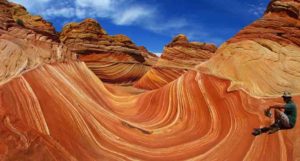
2. The Wave, Arizona
The Wave – Arizona
The Wave is a sandstone rock formation situated close its southern boundary with Utah in Arizona, United States. The structure is located on the Coyote Buttes hills in the Colorado Plateau’s Paria Canyon-Vermilion Cliffs Wilderness. The region is managed at the Grand Staircase-Escalante National Monument Visitor Center in Kanab, Utah by the Land Management Bureau (BLM).
Among hikers and photographers, the structure is well known for its colourful, ondulating shapes and the hard walk needed to achieve it and this is one of the best Places To Visit in United States of America. Because of the brittle nature of the training and the big amount of individuals wanting to attend it, the Kanab Visitor Center uses a regular lottery scheme to issue only ten licenses next day in individual. Furthermore, ten internet licenses are accessible four months prior to a scheduled journey for each date. Those who have acquired licenses will receive a map and data about the walk.
The Wave is made up of intersecting U-shaped troughs eroded into Jurassic age Navajo Sandstone. The two main stone forming troughs are 62 feet (19 m) wide by 118 feet (36 m) lengthy and 52 feet (16 m) broad by 7 feet (2 m). Initially, these troughs along joints within the Navajo Sandstone were eroded by rare runoff. The drainage basin, which supplied rainwater to these troughs, shrank after their creation to the stage that the runoff became inadequate to help cut these troughs.
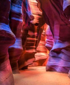
3. Antelope Canyon, Arizona
Antelope Canyon
Antelope Canyon is an American Southwest slot canyon. It’s on the west of Page, Arizona, Navajo territory. Antelope Canyon involves two distinct, picturesque canyon slot segments, separately referred to as ‘ Upper Antelope Canyon ‘ or ‘ The Crack ; ‘ and ‘ Lower Antelope Canyon ‘ or ‘ The Corkscrew. ‘
Tsé bighánílíní is the name Navajo for the Upper Antelope Canyon, which implies’ the location where water passes through trees.’ The Lower Antelope Canyon is the Navajo Parks and Recreation Department’s Hazdistazí (called “Hasdestwazi”), or’ spiral stone arches.’ Both are in the Navajo Nation’s LeChee Chapter. The canyons can only be accessed through a guided trip, This is a best place to feel happy and joy so it is one of best of Places To Visit in United States of America.
Navajo Sandstone erosion created the Antelope Canyon, mainly owing to flash flooding and secondary owing to other sub-aerial procedures. Rainwater flows into the expansive basin above the slot canyon segments, particularly during the monsoon season, picking up velocity and sand as it flows through the tight passages. The passages deteriorated over moment, deepening the hallways and brushing difficult corners in such a manner that distinctive “flowing” forms in the rock were formed.
4. Dry Tortugas National Park, Florida
Dry Tortugas National Park
Dry Tortugas National Park is about 68 miles (109 km) south of Key West in the Gulf of Mexico, a national park in the United States. The park retains Fort Jefferson and the seven islands of Dry Tortugas, the Florida Keys ‘ most remote and westernmost. The coral reefs of the archipelago are the least damaged of the reefs of Florida Keys.
The park is known for its abundant marine life, tropical bird breeding grounds, colourful coral reefs, shipwreck myths and sunken treasures. Fort Jefferson, a huge but incomplete coastal fortification, is the centerpiece of the park. Fort Jefferson is the Western Hemisphere’s biggest brick masonry structure, consisting of over 16 million bricks. Only Fort Monroe, Virginia, and Fort Adams, Rhode Island, exceed it in size among U.S. bases. Dry Tortugas is distinctive in its mixture with important historical artifacts of a mainly undisturbed tropical ecosystem.
The park is only accessible by seaplane or boat and has an average of approximately 63,000 visitors per year between 2008 and 2017. Snorkeling, picnicking, bird watching, hiking, scuba diving, fishing for saltwater and kayaking are some of the activities.
Dry Tortugas National Park is part of the Biosphere Reserve of Everglades & Dry Tortugas, established by UNESCO under its Man and Biosphere Program in 1976.
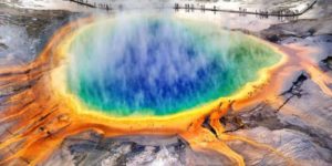
5. Yellowstone National Park, Wyoming, Montana, and Idaho
Yellowstone National Park is an American national park located in Wyoming, Montana, and Idaho.
Yellowstone National Park is a Wyoming, Montana, and Idaho American domestic park. It was set up by the U.S. Congress and President Ulysses S. Grant entered it into law on March 1, 1872. Yellowstone was the first national park in the U.S. and the first national park in the globe is also commonly kept. The park is renowned for its wildlife and numerous geothermal features, including Old Faithful Geyser, one of its most famous characteristics. It has many kinds of ecosystems, but the most common is the subalpine forest. It is component of the ecoregion of the South Central Rockies forests “Amazing places to visit in united states of america”.
Native Americans have resided for at least 11,000 years in the Yellowstone region. During the early-to-mid-19th century, apart from trips by mountain men, structured exploring did not start until the early 1860s. Originally, park management and control falls within the jurisdiction of the Interior Secretary, the first being Columbus Delano. But the U.S. Subsequently, the Army was commissioned to oversee Yellowstone management for a period of 30 years from 1886 to 1916.
Yellowstone is on the southeastern edge of the Snake River Plain, a large U-shaped curve through the hills stretching from Boise, Idaho about 400 miles (640 km) south.
Yellowstone volcanism is thought to be related to the Snake River plain’s somewhat earlier volcanism. Thus, Yellowstone is the active component of a hotspot that over time has shifted south. The source of this volcanic hotspot is controversial. One hypothesis argues that a mantle plume led the Yellowstone hotspot to migrate south, while another hypothesis explains migrating hotspot volcanism as a consequence of the fragmentation and dynamics of the subdued Farallon Plate in the middle of Earth.
The Yellowstone Caldera is North America’s biggest volcanic scheme. It was called a “supervolcano” because extremely big explosive eruptions created the caldera. It is projected that the magma chamber below Yellowstone is a single linked tank, approximately 37 miles (60 km) long, 18 miles (29 km) broad and 3 to 7 miles (5 to 12 km) depth.
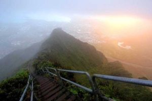
6. Haiku Stairs of Oahu, Hawaii
Haiku Stairs of Oahu, Hawaii
The Haalikū Stairs, also referred as the Heaven Stairway or Haalikū Ladder, is a challenging walking path on Hawaii’s island of Oalmahu. The total of 3,922 steps cover the mountain range of Oahu’s Ko’olau. “Haiku” refers not to the poetry genre of Japan. The region is named after the Kahili flower “Ha regikū.”
Contractors for the United States in 1942 Navy started the development of the Haferikū Radio Station, a top secret unit to be used to convey radio signals to Navy vessels then working across the Pacific. In order to obtain the necessary height for the antennae, they were stretched by the Navy across the natural amphitheater, Haifikū Valley. In addition to the metal steps, some remaining sections of the wooden ladder can still be seen.
7. Hamilton Pool, Texas
Hamilton Pool, Texas. Flickr: dawilson / Creative Commons
Hamilton Pool Preserve is a natural pool that was formed as a result of huge erosion thousands of years earlier when an underground river dome crashed. Around 23 miles (37 km) north of Austin, Texas off Highway 71, the pool is situated. Hamilton Pool has been a famous summer swimming place for tourists and inhabitants of Austin since the 1960s. Hamilton Pool Preserve is a preserved natural habitat of 232 acres (0.94 km2) with a jade green pool in which a 50-foot (15 m) waterfall flows with water temperatures exceeding 50 degrees Fahrenheit.
The pond is enclosed by big calcareous rocks that lie at the bottom of the water; big stalactites develop from the top of the roof. Moss, maidenhair fern and cliff swallows are close to the grotto’s roof and adjacent hills. The preserve’s Ashe juniper (cedar) uplands are home to the endangered warbler golden-cheeked.
There is no chemical treatment of the natural pool and creek, so water quality is constantly tracked and swimming is sometimes limited. Hamilton Pool is a protected area, part of the Balcones Canyonlands Preserve.
8. Horseshoe Bend, Arizona
Horseshoe Bend, Arizona
Horseshoe Bend is a Colorado River incised horseshoe meander close the city of Page, Arizona, USA, This is from the best places to visit in united states of america.
Horseshoe Bend is situated in the Glen Canyon National Recreation Area, about 4 miles (6.4 km) southeast of Page, 5 miles (8.0 km) upstream from the Glen Canyon Dam and Lake Powell.
It is available from a parking area near the U.S. by walking a 1.5-mile (2.4 km) round journey. Route 89 on the southwest side of Page.
From the steep cliff above, you can see Horseshoe Bend.
The view is 4,200 feet (1,300 m) above sea level, and the Colorado River is 3,200 feet (980 m) above sea level, resulting in a fall of 1,000 feet (300 m).
The lookout has recently become a significant place for tourists. By 2018, social media place mentions had prompted a significant rise in the amount of tourists.
9. Bryce Canyon, Utah
Bryce Canyon (Credit: Luca Galuzzi, CC by 2.5)
Bryce Canyon National Park is an American domestic park in southeastern Utah. The park’s main characteristic is Bryce Canyon, which is not a canyon despite its title, but a series of gigantic natural amphitheaters along the Paunsaugunt Plateau’s southern edge. Bryce is characteristic because of the geological structures called hoodoos, which are created by frost weathering and river and lake bed erosion of sedimentary rocks. The rocks ‘ red, orange, and white colours provide park tourists with amazing opinions. Bryce Canyon National Park is much lower than neighboring Zion National Park and lies at a much higher elevation. The rim at Bryce varies from 8,000 to 9,000 feet (2,400 to 2,700 m).
The area of Bryce Canyon was settled in the 1850s by Mormon pioneers and was named after Ebenezer Bryce, who in 1874 lived in the area. Originally appointed as a national monument by President Warren G. Harding in 1923, the region around Bryce Canyon was redesigned by Congress in 1928 as a national park.
The park includes 35,835 acres (55,992 sq mi; 14,502 ha; 145,02 km2) and gets significantly fewer tourists than Zion National Park (approximately 4.3 million in 2016) or Grand Canyon National Park (approximately 6 million in 2016), owing mainly to the more distant place of Bryce. Bryce Canyon got 2,365,110 recreational tourists in 2016, a 35 percent boost from the previous year.
10. Lake Tahoe, California/Nevada
Lake Tahoe, California/Nevada
Lake Tahoe in the U.S. Sierra Nevada is a big freshwater lake. Situated at 6,225 ft (1,897 m) south of Carson City, it straddles the state boundary between California and Nevada. Lake Tahoe is North America’s biggest alpine lake and passes only the five Great Lakes as the biggest by quantity in the United States at 122,160,280 acres (150.7 km3). Its elevation is 501 m (1,645 ft), making it the second longest in the United States after Oregon’s Crater Lake (1,949 ft or 594 m).
The lake was created as portion of the Lake Tahoe Basin about two million years ago, shaping the contemporary magnitude during the ice ages. It is renowned on all sides for the clearness of its water and the panorama of neighboring hills. Also known as Lake Tahoe, or merely Tahoe, is the region surrounding the lake. More than 75% of the lake’s catchment is domestic forest property, which includes the U.S. Forest Service’s Lake Tahoe Basin Management Unit so this is one of Places To Visit in United States of America.
Lake Tahoe is one of Nevada and California’s main tourist attractions. It is home to year-round winter sports, indoor summer recreation, and landscapes. Snow and ski resorts are a major component of the economy and reputation of the area. Several lakeside casino casinos are also available on the Nevada hand, with roads offering year-round access to the whole region.
11. Badlands National Park, South Dakota
Badlands National Park, South Dakota
Badlands National Park is a national park in the southwest of South Dakota. In addition to the biggest undisturbed mixed grass prairie in the United States, the park protects 242,756 acres (379.3 sq mi; 982.4 km2) of significantly eroded buttes and pinnacles. The park is managed by the National Park Service, with the South Unit co-managed by the tribe of Oglala Lakota, from the best places to visit in united states of america.
The Badlands Wilderness preserves the park’s 64,144 acres (100.2 sq mi; 259.6 km2) as a specified wildlife area, and is one location where the black-footed ferret, one of the world’s most endangered mammals, has been brought into the wild. The South Unit, or Stronghold District, includes sites of 1890s Ghost Dances, a former U.S. Air Force bomb and gunnery range, and Red Shirt Table, the highest point of the park at 1,020 m (3,340 feet).
It was not established until January 25, 1939, authorized as the Badlands National Monument on March 4, 1929. On November 10, 1978, Badlands was redesigned as a national park. The Ben Reifel Visitor Center was built for the memorial in 1957–58 under the Mission 66 scheme. The monument also administers the National Historic Site of the neighboring Minuteman Missile. Partially filmed in Badlands National Park were films such as Dances with Wolves (1990) and Thunderheart (1992).
12. Palouse Falls, Washington
Palouse Falls, Washington
The Palouse Falls are located on the Palouse River, approximately 4 miles (6 km) downstream of the Snake River confluence in southeast Washington, USA. The height of the drops is 198 ft (60 m). The hills comprise of an upper drop with a drop of about 20 ft (6.1 m), 1000 ft (305 m) north-northwest of the primary drop, and a smaller drop with a drop of 198 ft (60 m).
The canyon at the cliffs is 115 m (377 ft) profound, showing the Columbia River Basalt Group’s big cross-section. These hills and the upstream canyon are an significant characteristic of the channeled scablands formed during the Pleistocene era by the huge rains of Missoula that periodically spread across eastern Washington and across the plateau of the Columbia River is amazing Waterfall you must visit in Places To Visit in United States of America.
The ancient river Palouse flowed to the Columbia River through the presently dry Washtucna Coulee. The Palouse Falls and adjacent canyons were formed when the Missoula floods overturned the ancient Palouse River’s southern valley wall, diverting it by erosion of a fresh channel to the present route to the Snake River.
The region is distinguished by interconnected and suspended flood-created coulees, cataracts, diving basins, kolk-created potholes, rock benches, buttes, and typical scabulary pinnacles. Palouse Falls State Park is situated at the falls, preserving this unique portion of the picturesque region.
13. Nā Pali Coast State Park, Hawaii
Nā Pali Coast State Park, Hawaii
The Nā Pali Coast State Park is a Hawaiian national park of 6,175 acres (2,499 ha) situated in the middle of the hilly 16 miles (26 km) along Kauai’s southwest coast, the longest inhabited Hawaiian island. The coast of Nā Pali itself stretches southeast from Kee Beach to Polihale State Park. The na bari (elevated hills) rise above the Pacific Ocean as elevated as 4,000 feet (1,200 m) along the coast. To safeguard the Kalalau Valley, the State Park was created.
The Hono O Nā Pali State Natural Reserve is located to the south of the national park. It was set up in 1983 and expanded in 2009 to more than 3,578 acres (14.5 km2). Hiking paths and highways for visitors in Waimea Canyon have access to the steep slopes from Koke’e Road (route 550).
14. Glacier National Park, Montana
Glacier National Park
Glacier National Park is an American national park situated on the Canada-U.S. border in southeastern Montana, close to Alberta and British Columbia regions of Canada. The park covers more than 1 million acres (4,000 km2) and contains sections of two mountain ranges (the Rocky Mountains sub-ranges), more than 130 named lakes, more than 1,000 distinct plant species, and hundreds of animal species. This vast untouched ecosystem is at the center of what has been called the “Crown of the Continent Ecosystem,” an area of 16,000 square miles (41,000 km2) of protected territory.
Native Americans first populated the region that became the Glacier National Park. When European explorers arrived, it was dominated in the east by the Blackfeet and in the west by the Flathead. Under stress, the Blackfeet surrendered to the federal government in 1895 the hilly sections of their treaty lands; it subsequently became component of the park. Soon after the park was set up on May 11, 1910, the Great Northern Railway built a range of accommodations and chalets.
15. Thor’s Well, Oregon
Thor’s Well, Oregon
There is a natural cave on the shore of Oregon that seems to drain the ocean.
At high tide, or during floods, the place is most impressive when water washes violently over the rocks and funnels into the cave. Unattended tourists to the site run the danger of being thrown straight into the maelstrom during these sudden torrents. Photographers and nature enthusiasts proceed to flock to the magnificent fountain, regardless of the hazards, to see the hostile waters that live up to the stormy character of their godly namesake. The best time to check Thor’s Well is to watch it without water one hour before the high tide and then watch the structure fill up.
16. Sequoia National Park, California
Sequoia National Park
Sequoia National Park in the central Sierra Nevada south of Visalia, California, is an American national park. The reserve was created on September 25, 1890 to safeguard 404,064 acres of wooded hilly land (631 sq mi; 163,519 ha; 1,635 km2). The park includes the highest point in the contiguous United States, Mount Whitney, at 14,505 feet (4,421 m) above sea level, covering a vertical relief of nearly 13,000 feet (4,000 m).
The park is north of and adjacent to Kings Canyon National Park ; the two areas are administered as Sequoia and Kings Canyon National Parks by the National Park Service together. The Sequoia-Kings Canyon Biosphere Reserve was named by UNESCO in 1976.
The park is noteworthy for its giant sequoia forests, including the General Sherman tree, Earth’s biggest tree. In the Giant Forest, which includes five of the world’s ten largest trees, the General Sherman tree develops. The Giant Forest is linked to General Grant Grove, home of the General Grant tree among other giant sequoias, by the Generals Highway to Kings Canyon National Park.
17. Niagara Falls, New York
Niagara Falls, New York
Niagara Falls is a town in the county of Niagara, New York, USA. The town had a complete population of 50,193 as of the 2010 census, down from the 55,593 reported in the census of 2000. It is close to the Niagara River, across from Niagara Falls City, Ontario, and named after the well-known Niagara Falls they share. The town is located within the Metropolitan Statistical Area of Buffalo–Niagara Falls and the West New York area, This is from amazing places to visit in united states of america.
While the town was formerly occupied by Native Americans, Europeans who moved in the mid-17th century to the Niagara Falls started opening up companies and developing infrastructure. Scientists and entrepreneurs later started to harness the capacity of the Niagara River for electricity in the 18th and 19th decades, and the town started to attract producers and other companies attracted by the offer of cheap hydroelectric power. However, following an effort at urban renewal under then-Mayor Lackey, the town and region experienced an financial decrease after the 1960s.
18. Great Smoky Mountains, North Carolina/Tennessee
Great Smoky Mountains, North Carolina/Tennessee
The Great Smoky Mountains are a hill range that rises in the southern United States along the Tennessee–North Carolina boundary. They are an Appalachian Mountains sub-range and are component of the Physiographic Province of the Blue Ridge. Sometimes the range is called the Smoky Mountains and the name is usually abbreviated to the Smokies. The Great Smokies are best regarded as the headquarters of the National Park of the Great Smoky Mountains, protecting most of the area. Established in 1934, the park is the most visited national park in the United States with over 11 million visits per year.
The Great Smokies are component of a Biosphere Reserve International. The range is home to an approximately 76,000 ha (187,000 acres) of old growth forest, the biggest such cluster to be found west of the Mississippi River. One of the most varied ecosystems in North America is the cove hardwood forests in the reduced altitudes of the range, and the Southern Appalachian spruce-fir forest that covers the upper parts of the range is the biggest of its kind. The Great Smokies are also home to the Eastern United States ‘ densest black bear population and the most varied salamander community outside the tropics.
19. Northern Lights, Alaska
Northern Lights, Alaska
While many tourists believe they must go to Europe to see the Northern Lights, you can truly see this phenomenon without abandoning the United States. There are many Americans heading for the opportunity to see the aurora borealis in northern Alaska. It may be cold in winter (temperatures may drop to-30 ° F), but coastal Alaskan Arctic— where skies tend to be clearer— is one of the world’s greatest locations to see this renowned light display.
20. Carlsbad Caverns, New Mexico
Carlsbad Caverns, New Mexico
Carlsbad Caverns National Park is an American National Park in southern New Mexico’s Guadalupe Mountains. The park’s main attraction is the Carlsbad Cavern show cave. Visitors to the cave can walk or bring an elevator from the visitor center on their own through the natural door.
On US Highway 62/180, about 18 miles (29 km) southeast of Carlsbad, New Mexico, the park gate is situated. The National Park of Carlsbad Caverns is involved in the Junior Ranger Program. The reserve has two listings on the Historic Places National Register: the Historic District of Caverns and the Historic District of Rattlesnake Springs. Approximately two-thirds of the park has been laid aside as a wilderness area to help guarantee that future habitat modifications are not created.
Carlsbad Cavern contains a big room of calcareous, merely called the Big Room, which is nearly 4,000 feet (1,220 m) long, 625 feet (191 m) broad, and at its lowest level, 255 feet (78 m). The Big Room is North America’s third biggest chamber and the world’s twenty-eighth biggest.
This image shows the divisions between Earth’s layers. The ancient, continent-sized rock regions encircle the liquid outer core. Credit: Lawrence Livermore National Laboratory
Ancient, distinct, continent-sized regions of rocks, isolated since before the collision that created the Moon 4.5 billion years ago, exist hundreds of miles below the Earth’s crust, offering a window into the building blocks of our planet, according to new research.
The new study in the AGU Journal Geochemistry, Geophysics, Geosystems used models to trace the location and origin of volcanic rock samples found throughout the world back to two solid continents in the deep mantle. The new research suggests the specific giant rock regions have existed for 4.5 billion years, since Earth’s beginning.
Previously, scientists theorized that separated continents in the deep mantle came from subducted oceanic plates. But the new study indicates these distinct regions may have been formed from an ancient magma ocean that solidified during the beginning of Earth’s formation and may have survived the massive Moon-creating impact.
Determining the masses’ origin reveals more details about their evolution and composition, as well as clues about primordial Earth’s history in the early Solar System, according to the study’s authors.
It’s amazing that these regions have survived most of Earth’s volcanic history relatively untouched, said Curtis Williams, a geologist at the University of California, Davis, in Davis, California and lead author of the study.
Looking inward
The mantle is a layer of rock, stretching 2,900 kilometers (1,802 miles) down inside the Earth. Earth’s molten, liquid, metallic core lies beneath the mantle. The core-mantle boundary is where the solid mantle meets the metallic liquid core.
Scientists knew from past seismic imaging studies that two individual rock bodies existed near the core-mantle boundary. One solid rock body is under Africa and the other is under the Pacific Ocean.
Seismic waves, the vibrations produced by earthquakes, move differently through these masses than the rest of the mantle, suggesting they have distinct physical properties from the surrounding mantle. But geologists couldn’t determine whether seismic waves moved differently through the core-mantle continents because of differences in their temperature, mineral composition or density, or some combination of these properties. That meant they could only hypothesize about the separate rocky masses’ origin and history.
“We had all of these geochemical measurements from Earth’s surface, but we didn’t know how to relate these geochemical measurements to regions of Earth’s interior. We had all of these geophysical images of the Earth’s interior, but we didn’t know how to relate that to the geochemistry at Earth’s surface,” Williams said.
Primitive material and plumes
Williams and his colleagues wanted to determine the distinct masses’ origin and evolution to learn more about Earth’s composition and past. To do this, they needed to be able to identify samples at Earth’s surface with higher concentrations of primitive material and then trace those samples back to their origins.
Scientists often take rock samples from volcanic regions like Hawaii and Iceland, where deep mantle plumes, or columns of extremely hot rock, rise from the areas near the core, melt in the shallow mantle and emerge far from tectonic fault lines. These samples are made of igneous rock created from cooling lava. The study’s authors used an existing database of samples and also collected new samples from volcanically active areas like the Balleny Islands in Antarctica.
Geologists can measure specific isotopes in igneous rocks to learn more about the origin and evolution of the Earth. Some isotopes, like Helium-3, are primordial, meaning they were created during the Big Bang. Rocks closer to Earth’s crust have less of the isotope than rocks deeper underground that were never exposed to air. Samples with more Helium-3 are thought to come from more primitive rocks in the mantle.
The researchers found some of the samples they studied had more Helium-3, indicating they may have come from primitive rocks deep in the Earth’s mantle.
The researchers then used a new model to trace how these primitive samples could have gotten to the Earth’s surface from the mantle. Geological models assume plumes rise vertically from deep within the mantle to the Earth’s surface. But plumes can move off course, deflected, due to various reasons. The new model took into account this plume deflection, allowing the study’s authors to trace the samples back to the two giant masses near the core-mantle boundary.
The combination of the isotope information and the new model allowed the researchers to determine the composition of the two giant masses and theorize how they may have formed.
Understanding the composition of specific rock masses near the core-mantle boundary helps geologists conceptualize ancient Earth-shaping processes that led to the modern-day mantle, according to the study’s authors.
“It’s a more robust framework to try and answer these questions in terms of not making these assumptions of vertically rising material but rather to take into account how much deflection these plumes have seen,” Williams said.
Reference:
C. D. Williams et al. Primitive Helium Is Sourced From Seismically Slow Regions in the Lowermost Mantle, Geochemistry, Geophysics, Geosystems (2019). DOI: 10.1029/2019GC008437
Note: The above post is reprinted from materials provided by American Geophysical Union. The original article was written by Abigail Eisenstadt .