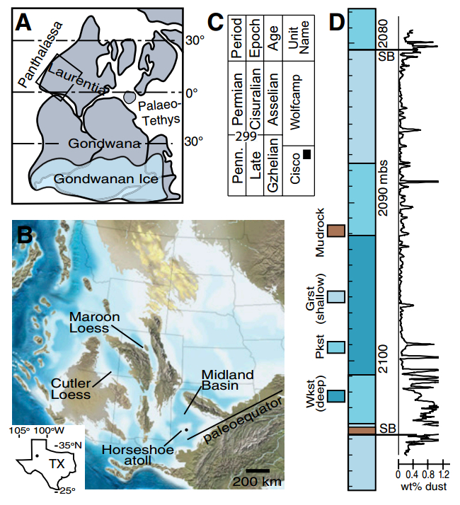
Figure 1 from Sur et al.: Late Paleozoic Pangaea, showing (boxed) region of interest in western tropical Pangaea.
Dust begets life, and Earth’s atmosphere 300 million years ago was perhaps the dustiest of all time, with large consequences for carbon cycling and the climate system. In a new paper for Geology, Sohini Sur and colleagues examine the bioavailability of iron in dust from Earth’s penultimate icehouse of the late Paleozoic. Dust links to carbon because of the iron — a key nutrient for nearly all life, so atmospheric dust acts as a fertilizer.
Vast volumes of dust deposits dating from the late Paleozoic, from both land areas and marine reefal deposits, record a remarkably dusty atmosphere. Moreover these dust deposits contain unusually high concentrations of reactive iron. This concomitance — dust with exceptionally high values of bioavailable iron — implies major ecosystem fertilization and an associated massive drawdown of atmospheric carbon.
The biogeochemical impacts of iron-rich dust to the oceans are known for Earth’s recent record but unexplored for deep time, despite recognition of large ancient dust fluxes, particularly during the late Paleozoic. Sur and colleagues report a unique iron relationship for Upper Pennsylvanian mudrock of eolian origin that records lowstand (glacial) conditions within a carbonate buildup of western equatorial Pangaea (now the western United States).
With iron fertilization proposed as a serious geoengineering scheme to control future atmospheric carbon and attendant climate change, authors Sur and colleagues write, “It behooves us to study the consequences of analogous events as archived in Earth’s deep-time record.”
Reference:
Sohini Sur, Jeremy D. Owens, Gerilyn S. Soreghan, Timothy W. Lyons, Robert Raiswell, Nicholas G. Heavens, Natalie M. Mahowald. Extreme eolian delivery of reactive iron to late Paleozoic icehouse seas. Geology, 2015; G37226.1 DOI: 10.1130/G37226.1
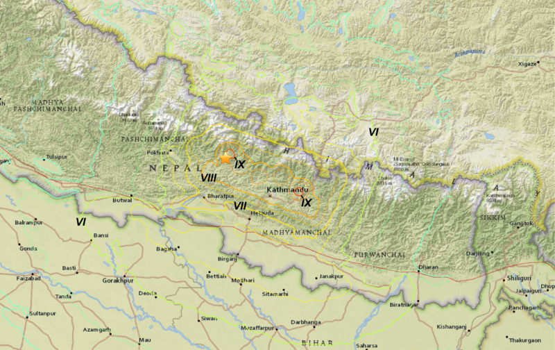
Isoseismal map for the 2015 Gorkha earthquake, showing contours of seismic intensity on the Mercalli scale Credit: USGS
Researchers from the University of Southampton are using mobile phone data to monitor the movement of people affected by the earthquake in Nepal and help with the continuing relief effort in the country.
Just a week after the disaster took place, the team, working as part of the WorldPop project and Flowminder Foundation, began daily monitoring of anonymised phone data to track the displacement of populations in the affected areas. Five months on, they are continuing to supply accurate, up-to-date reports to the United Nations.
Flowminder Director and Professor of Geography at Southampton Andy Tatem says: “We had been planning to conduct a project of this kind in Nepal since 2014 and the Flowminder team was already in Kathmandu setting this up. As a result, when the quake struck, we were able to respond rapidly and were well-placed to help.
“Although we have used mobile phone data before to measure population movements, this is the first time we have used the method in an ongoing ‘live’ situation. By watching how the population moves on a daily basis, we have been able to help directly with aid efforts and the rebuilding of infrastructures.”
Every time a person uses a mobile it sends information to a receiving tower and gives an approximate location of where they are. When this information is repeated multiple times, over millions of users, a detailed picture can be extracted of population density, movement and how it changes over time in a given area.
In Nepal, the researchers linked up with the largest mobile phone operator Ncell to analyse anonymised data from 12m mobiles across the country. By comparing information on the movement of these phones after the quake, with population distributions and movements before it hit, the team has established trends of where people are moving to and from. The work to monitor this continues.
The Nepal earthquake hit on 25 April 2015 killing over 9,000 people and injuring more than 23,000 – with a magnitude of 7.8Mw1. It was followed by more than 300 aftershocks. An estimated 500,000 buildings were destroyed or severely damaged. Economic losses have been estimated at $7,065m.
Two weeks after the quake, the researchers’ data showed that an estimated extra 500,000 people had left Kathmandu Valley (taking into account normal patterns of movement). Most went to the surrounding districts and the Terai areas in the south and southeast of Nepal – something which had previously been completely unknown. On a wider scale, across the country an estimated 1.8m people left their home district.
Latest analysis shows that of those people who left their homes soon after the disaster, most have now returned – with approximately four to 14 per cent still remaining elsewhere. Return rates have varied across districts of Nepal, with those from Kathmandu Valley the slowest to return. Kathmandu city centre has seen the largest inflow of population.
Dr Robin Wilson of Geography and Environment at Southampton, who led analyses of the phone data, comments: “Large population movements occur each time there is a natural disaster and there is often limited information to help understand where affected people move to—making it difficult to plan a response. The use of mobile phone data that we have pioneered has now proved to be invaluable in a real scenario—helping to get support to where it is needed in an effective way.”
According to new U.S. Geological Survey research published today in the Proceedings of the National Academy of Sciences, springs and marshes in the desert outside Las Vegas expanded and contracted dramatically in response to past episodes of abrupt climate change, even disappearing altogether for centuries at a time when conditions became too warm. This new record, gleaned from dirt and rocks exposed in the desert just outside the city limits, provides an unprecedented look into how climate change can affect fragile desert ecosystems in the American Southwest.
Kathleen Springer, a geologist with the USGS and former Senior Curator at the San Bernardino County Museum, was the principal investigator and lead scientist for this study showing that desert wetlands are extremely sensitive to climate change.
“This is a story of water,” said Springer. “Water was plentiful in the desert at times in the past, but when climate warmed, springs and wetlands dried up, and the plants and animals living in the harsh desert environment were out of luck.”
During the Pleistocene, between approximately 100,000 and 10,000 years ago, wetlands dotted the landscape in the area just north of Las Vegas, attracting a plethora of ice age animals, including mammoths, sloths, sabre-toothed cats, dire wolves, and extinct species of bison, horse, and camel, and later, the first human inhabitants to the area.
Today, existing desert wetlands are home to a number of threatened and endangered species that rely on the ecosystem for water in an otherwise arid landscape. Their fate may lie in the hands of a rapidly changing climate.
“What we’re seeing in the geologic record frames what we are observing today,” said Springer. “The drought that California is currently experiencing is extreme, but droughts are an inherent part of the climate system and have occurred repeatedly in the past.”
The study was initiated by the Bureau of Land Management, which called for an integrative approach to studies that emphasize the geological age and context of fossils, as well as a comprehensive analysis of how local hydrologic systems responded to climate change in the past.
“Scientists collect fossils all the time,” said Scott Foss, a senior paleontologist with the BLM. “What is remarkable about this work is the vision that Kathleen had of making sure her team understood the intricacies of the deposits in incredible detail, which allowed them to determine how climate affected the local landscape. It was an immense undertaking, and one that will serve as a benchmark for generations to come for those interested in understanding the effects of climate change on desert ecosystems.”
Studies examining the effects of climate change on springs and desert wetlands will continue through the USGS’s Climate and Land Use Change Research and Development Program, and will build upon the investigations conducted in the Las Vegas Valley, a large portion of which is now protected as Tule Springs Fossil Beds National Monument.
When the monument was established in December 2014, the BLM turned their stewardship over to the National Park Service, who will determine how to interpret the unique land and its former inhabitants for the public.
“The future of this newly designated national monument and what it can tell us about the effects of climate change is all about the past,” said Springer. “And the past is the key to the present.”
Reference:
K. B. Springer et al. Dynamic response of desert wetlands to abrupt climate change, Proceedings of the National Academy of Sciences (2015). DOI: 10.1073/pnas.1513352112
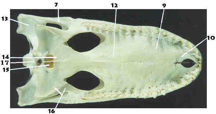
Those holes towards the back, labelled by I7, are the choanae.
Crocodiles are freakin’ amazing animals. They’ve been around for about 250 million years, and throughout this time have survived two mass extinctions, and at least twice decided to hitch up and take to the seas. Their historical diversity, and general weirdness, was vast compared to what we see in modern crocs, which are on the face of it all fairly similar. Extinct forms included those that looked like armadillos and even ate plants, as well as some that became gigantic and streamlined for swimming out to sea. Others were up to 12 metres long, and snacked on dinosaurs!
All modern crocs, alligators, caimans, and gharials belong to a group known as Crocodylia. The origins of this group can be traced back to the Cretaceous, when many of these bizarre croc-cousins, known collectively as crocodyliforms, where still around. Trying to work out the evolutionary origins of modern crocs though has proven to be a bit confusing for palaeontologists. Part of this is simply due to the fact that the fossil record preserves incomplete remnants of the lineage leading to modern crocs, which in turn creates issues in our understanding the relationships and anatomical changes that led to the origin of Crocodylia.
One thing we do know is that a group known as Eusuchia are the direct ancestors of modern crocs – Crocodylia belongs to Eusuchia, but not all eusuchians are crocodylians, if that makes sense. That’s because some eusuchians went extinct during the Cretaceous, leaving just crocodylians (and a couple of other non-eusuchian groups like the now extinct marine dyrosaurids) around to take charge. One of the problems which croc workers have been trying to figure out is what defines Eusuchia, and therefore what croc species can be assigned to this group. If we know this, then we can look at the evolutionary changes that led to the origins of modern crocodilians, and why these chappies became so successful.
Eusuchians have been traditionally recognised based on a couple of really important modifications to the ‘standard’ crocodyliform skeleton that reflect major changes in their lifestyle. One of these involves the movement of the choanae, an opening in the top of the mouth that helped crocs to breathe more efficiently, from a position closer to the nostrils to a position further back in the skull. This was due to the development of what’s called the secondary palate, the bony surface in the roof the mouth which grew as the overall skull lengthened in crocs to form the snout. Another important development of eusuchians was to do with the vertebrae. Until eusuchians, crocodyliforms (remember, the ancestors of modern crocs) had vertebrae in which the articular surfaces were either flat or concave, which limited mobility of the vertebral column. In Eusuchia, the articular surface facing towards the tail became progressively more hemispherical-shaped, or convex outwards, to what we call a ‘procoelous condition’, forming a sort of ball and socket articulation. This would have allowed greater flexibility of the vertebral column, which is a pretty useful adaptation to have.
So why the confusion about what the origins of Eusuchia? Well, for starters, a lot of fossils that look like they could be a eusuchian are often preserved in a way that we can’t tell what the choanae and vertebral columns looked like, or these bits are just missing. This leads to quite a lot of uncertainty about what constitutes a ‘true’ eusuchian, and has complicated both the species that can be assigned to Eusuchia, and the pattern of acquisition of these important anatomical features. Recently, a couple of papers by Alan Turner overhauled Eusuchia, and he suggested that other groups, including Paralligatoridae and Atoposauridae could both be included within Eusuchia too. However, I don’t think this is 100% correct, as few if any of the species from these groups can be conclusively shown to have the features that define Eusuchia as mentioned above, and it is possible that atoposaurids and paralligatorids lie outside of Eusuchia (disclosure: I have a paper in review discussing this a bit at the moment). So that’s a nice additional layer of confusion to add in!
So that’s a whole lot of background, and I think important to wrap our heads around for a couple of reasons. Firstly, it shows that trying to figure out the taxonomy and evolutionary relationships of extinct animals is complicated, and pretty dynamic as far as what constitutes science (evidence-based inference) goes. Secondly, it shows how complicated our current understanding of the origins of modern crocs is, and the reasons for this complexity. Thirdly, it highlights how important new fossil finds might be in helping to unravel some of this evolutionary mess, which provides us with a nice segue into new croc species klaxon.
Well, actually, two new crocs! A new study in PLOS ONE has identified two new species of crocodyliform from the same genus, Loheucosuchus (Low-hay-kwo-soo-kus). The first of these new crocs comes from near the village of Fuentes, Cuenco, in Spain, from a fossil locality known as Lo Hueco. The fossils here come from a time right towards the end of the Cretaceous, in time intervals known as the Campanian and Maastrichtian. This new species was called Lohuecosuchus megadontos, and it’s probably pretty obvious where the genus name comes from. The species name means ‘big tooth’, and refers to the well, uniquely big teeth this new croc has! ‘suchus’ is Latinized from the Greek word souchos, and refers to an Egyptian crocodile-headed god!
As well as this new genus and species, they named a second new species referred to the new genus, Lohuecosuchus mechinorum, from the Fox-Amphoux site from Department of Var in France, and based on extensive comparisons with previously known material referred to a different species. The species name ‘mechinorum’ in this case is from the Mechin Collection (in honour of Patrick and Annie Mechin) at the Muséee des Dinosaures in Espéraza, France, which houses the specimens.
These new findings seem to provide a bit of insight into how Late Cretaceous crocs from Europe are related. They all fit within a newly resolved group known as Allodaposuchidae, named after Allodaposuchus as is common when naming these types of group. Allodaposuchus has been known for quite a while from multiple localities referred to several species from the Late Cretaceous of Europe. It’s what we like to call in palaeontology a ‘taxonomic nightmare’.
Allodaposuchidae seems to be related to another group of fairly unusual crocs known as Hylaeochampsidae, named after (you guessed it) Hylaeochampsa, another croc known from the Early Cretaceous of the Isle of White in Europe. Now Hylaeochampsidae is a bit of a taxonomic mess. Previously, species from the Cretaceous of North America (known as Pachycheilosuchus) and another from Italy called Pietraroiasuchus (I can’t say it either..), where assigned to this group, along with others from Europe like Acynodon and Iharkutosuchus. But membership has always been in a bit of a state of flux, depending on which researchers you ask. This is important as historically, hylaeochampsids have been regarded as the earliest, or most basal, eusuchians. Solve Hylaeochampsidae, solve Eusuchia. Oh yeah, it’s all coming together now.
Importantly, this new study finds both Allodaposuchidae and Hylaeochampsidae together to be the sister group to Crocodylia. In non-phylogenetics speak, this means that these groups are the closest relatives to the group that includes all modern crocodiles, alligators, and gharials, with the three of them together sharing a common ancestor (i.e., common origin). Hylaeochampsidae is found to comprise just Hylaeochampsa, Acynodon, and Iharkutosuchus – three exclusively European crocs. This is important, as it pretty much cements the idea that Crocodylia originated in Europe from an exclusively European stock of eusuchian crocs. Or so it would seem..
However, I wouldn’t be a croc palaeontologist if I didn’t raise a few potential issues. Or at least, things that spring to mind. The way in which palaeontologists analyse the relationships of organisms is though what we call phylogenetic analysis. These produce ‘phylogenies’, commonly depicted as trees, which illustrate the hierarchical relationships of organisms. These analyses are based on data matrices that comprise the morphology of organisms reduced to numerical codes that describe different aspects of their anatomy, and the different conditions these can take across all animals considered. What this means is that often when designed, these character matrices are created to test very explicit hypotheses about organismal relationships, based on whatever it is you want at the time, such as the relationships of a group or the position of a particular animal (taxon). But what a lot of researchers do, I imagine mostly for convenience, is to take data matrices used to test previous hypotheses, and simply add a new species into that matrix to test what is by default a very different hypothesis. And that’s what happened here. The new study uses a matrix by Chris Brochu and Glenn Storrs, published back in 2012, designed to test the relationships of a new crocodylian species from the Pliocene-Pleistocene (the last few million years) of Kenya. So the question is, is that matrix adequate to test the relationships of a ‘basal eusuchian’ from the Late Cretaceous of Europe? By using a matrix designed to test the relationships of more advanced crocodylians, the character matrix will contain a lot more characters (anatomical features) that are found in more advanced crocodylians in order to resolve their relationships. By extension, this means that fewer of these characters will be appropriate to test ‘deeper’ crocodylian relationships back in the Cretaceous, and might explain why several species previously regarded as eusuchians are falling outside of this group in their analyses. If you think about the logic behind this, it’s like looking just at modern birds, and trying to figure out what the relationships of Archaeopteryx are from it. You have to sample much deeper from down in the tree at older forms more closely related to the target animal in order to adequately test its relationships. While I don’t think this is a major issue with the results and placement of Lohuecosuchus, and the resolution of the new group Allodaposuchidae, I think it would have been really good to test alternative relationships for it by using different and possibly more appropriate matrices.
As well as this, such potential inadequacy might help to explain a few of the oddities in their results. As well as just using the matrix of Brochu and Storrs, they added several taxa mentioned above to this matrix and ‘coded’ them for their morphology. These included Shamosuchus, Pietraroiasuchus, and Pachycheilosuchus, and which the new analysis found all to be outside of Eusuchia. Weird that. While perhaps not unexpected for anyone familiar with these crocs, it is probably due to the issues mentioned above, and not sampling other crocs from deeper down in the tree related to these. In addition, the use of the closely related Bernissartia as what we call an outgroup (the taxon used to define the sequence of morphological evolution by being the most ‘basal’ in the analyses) is probably not appropriate, as typically more distantly related taxa are needed in order to understand what the actual ‘basal’ features of a group are. This issue has been raised recently with crocs, which found a completely different placement for a major marine radiation known as Thalattosuchia to be in a different phylogenetic placement depending on what is used as an outgroup.
But, if the resolution of an allodaposuchid-hylaeochampsid only Eusuchia is true (along with Crocodylia), then it has some pretty important implications. Both of these groups went extinct at the end of the Cretaceous, in the mass extinction that also took out the pterosaurs, marine reptiles, and the non-avian dinosaurs. Could it be that this removed competition with early crocodylians, and allowed them to radiate in their absence? This supports recent studies which showed that crocs actually seemed to do pretty well after the end-Cretaceous mass extinction, and shows that while we might think of extinction as generally bad, it really depends on whether you’re one of the survivors or not..
So for now, I’d still say we still haven’t fully resolved Eusuchia, and the results of this new study should be taken with a pinch of salt. Still, a cool new croc, and I look forward to seeing future analyses including it to see where it fits within the broader scheme of croc evolution.
Reference:
Iván Narváez et al. New Crocodyliforms from Southwestern Europe and Definition of a Diverse Clade of European Late Cretaceous Basal Eusuchians, PLOS ONE (2015). DOI: 10.1371/journal.pone.0140679
Alan H. Turner et al. A Review of Shamosuchus and Paralligator (Crocodyliformes, Neosuchia) from the Cretaceous of Asia, PLOS ONE (2015). DOI: 10.1371/journal.pone.0118116
Christopher A. Brochu et al. A giant crocodile from the Plio-Pleistocene of Kenya, the phylogenetic relationships of Neogene African crocodylines, and the antiquity of in Africa , Journal of Vertebrate Paleontology (2012). DOI: 10.1080/02724634.2012.652324
Alan H. Turner et al. The monophyly of Susisuchidae (Crocodyliformes) and its phylogenetic placement in Neosuchia, PeerJ (2015). DOI: 10.7717/peerj.759
Note: The above post is reprinted from materials provided by Public Library of Science. This story is republished courtesy of PLOS Blogs: blogs.plos.org.
An international research team led by a scientist at New Zealand’s University of Otago has resolved the mystery of the processes involved in the Mediterranean Sea drying up around 5.6 million years ago.
The event, known as the Messinian Salinity Crisis (MSC), saw the Mediterranean become a 1.5km deep basin for around 270,000 years. It also left a kilometers-deep layer of salt due to seawater evaporation.
The cause of the MSC has been the subject of vigorous scientific debate, but now an international team of researchers led by Otago geologist Dr Christian Ohneiser have used a multidisciplinary approach to solve the puzzle.
Their findings are published this week in the prestigious international journal Nature Communications.
After the MSC was discovered in the 1970s, the early theory was that movements of the African, Arabian and Eurasian tectonic plates had led to the Mediterranean becoming landlocked. Later on, other scientists suggested that it was a global fall in sea levels due to growing ice sheets that cut the sea off from the Atlantic Ocean.
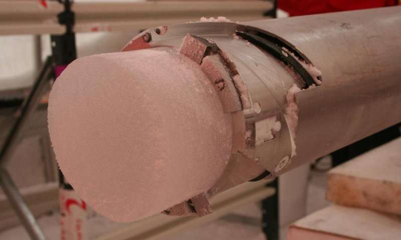
Dr Ohneiser and colleagues examined 60 sedimentary drill cores from around the edge of Antarctica.
“We determined that the continent’s ice was indeed growing in the lead up to the MSC, but the timings of key events did not precisely match,” Dr Ohneiser says.
The researchers then used a computer model of the Earth that simulated growth in the Antarctic ice sheet to see what geophysical impacts this would have aside from generally lowering the sea level.
“We found that the Antarctic ice sheet had an uneven effect on the global sea level because its growth resulted in a complex interplay between gravitational and rotational effects and the deformations to the Earth’s crust caused by ice advance and retreat,” he says.
The researchers determined that as the Mediterranean Sea evaporated, the Earth’s crust around the Strait of Gibraltar began rising up because the overlying load from the water was removed.
“This kept the Mediterranean isolated from the Atlantic Ocean until the crust began to relax and sink. At the same time, Antarctica began to melt, raising sea levels again.”
By around 5.33 million years ago, the rising sea level was just enough to wash over the thin land bridge at Gibraltar, resulting in a catastrophic flood that refilled the sea, he says.
Other researchers have previously shown that this deluge, known as the Zanclean flooding event, took only a few years to fully replenish the Mediterranean.
Dr Ohneiser says that one of the key implications of the study is that changes in global sea-level are uneven when ice sheets expand or retreat.
“Future melting of the large Southern or Northern hemisphere ice masses will result in an uneven rise in sea-level around the world, and this should be factored into future climate change scenarios,” he says.
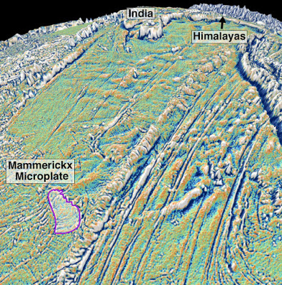
The newly discovered tectonic plate, named Mammerickx Microplate. Credit: University of Sydney
An international team of scientists has discovered the first oceanic microplate in the Indian Ocean—helping identify when the initial collision between India and Eurasia occurred, leading to the birth of the Himalayas.
Although there are at least seven microplates known in the Pacific Ocean, this is the first ancient Indian Ocean microplate to be discovered. Radar beam images from an orbiting satellite have helped put together pieces of this plate tectonic jigsaw and pinpointed the age for the collision, whose precise date has divided scientists for decades.
Reported in Earth and Planetary Science Letters, the team of Australian and US scientists believe the collision occurred 47 million years ago when India and Eurasia initially smashed into each other.
Researchers led by the University of Sydney School of Geosciences discovered that crustal stresses caused by the initial collision cracked the Antarctic Plate far away from the collisional zone and broke off a fragment the size of Australia’s Tasmania in a remote patch of the central Indian Ocean.
The authors, comprising Professor Dietmar Müller and Dr Kara Matthews from the University of Sydney and Professor David Sandwell from the Scripps Institution of Oceanography, have named the ancient Indian microplate the Mammerickx Microplate, after Dr Jacqueline Mammerickx, a pioneer in seafloor mapping.
The Mammerickx Microplate rotation is revealed by a rotating pattern of grooves and hills that turn the topography of the ocean floor into a jagged landscape. These so-called “abyssal hills” record a sudden increase in crustal stress, dating the birth of the Himalayan Mountain Range to 47 million years ago.
The ongoing tectonic collision between the two continents produces geological stresses that build up along the Himalayas and leads to numerous earthquakes every year—but this latest finding indicates how stressed the Indian Plate became when its northern edge first collided with Eurasia.
The new research shows that 50 million years ago, India was travelling northwards at speeds of some 15 centimetres a year—close to the plate tectonic speed limit. Soon after it slammed into Eurasia crustal stresses along the mid-ocean ridge between India and Antarctica intensified to breaking point. A chunk of Antarctica’s crust broke off and started rotating like a ball bearing, creating the newly discovered tectonic plate.
The discovery was made using satellite radar beam mapping from space, which measures the bumps and dips of the sea surface caused by water being attracted by submarine mountains and valleys, combined with conventional marine geophysical data.
Lead author Dr Matthews explains: “The age of the largest continental collision on Earth has long been controversial, with age-estimates ranging from at least 59 to 34 million years ago.
“Knowing this age is particularly important for understanding the link between the growth of mountain belts and major climate change.”
Co-author Professor Müller said: “Dating this collision requires looking at a complex set of geological and geophysical data, and no doubt discussion about when this major collision first started will continue, but we have added a completely new, independent observation, which has not been previously used to unravel the birth of this collision.
“It is beyond doubt that the collision must have led to a major change in India’s crustal stress field—that’s why the plate fragmentation we mapped is a bit like a smoking gun for pinning down the collision age.”
Co-author Professor Sandwell from the Scripps Institution of Oceanography said humans had explored and mapped remote lands extensively but the same was not true for our ocean basins.
“We have more detailed maps of Pluto than we do of most of our own planet because about 71 per cent of the Earth’s surface is covered with water,” Professor Sandwell said.
“Roughly 90% of the seafloor is uncharted by ships and it would take 200 ship-years of time to make a complete survey of the deep ocean outside continental shelves, at a cost of between two- to three billion US dollars.
“That’s why advances in comparatively low-cost satellite technology are the key to charting the deep, relatively unknown abyssal plains, at the bottom of the ocean.”
The paper ‘Oceanic microplate formation records the onset of India-Eurasia collision’ was be published in Earth and Planetary Science Letters last week.
Reference:
The paper ‘Oceanic microplate formation records the onset of India–Eurasia collision’ was be published in Earth and Planetary Science Letters last week. DOI:10.1016/j.epsl.2015.10.040
A UC Riverside-led study finds most aerosol species will increase under climate change associated with greenhouse-gas-induced warming. Credit: taylorandayumi.
Aerosols, tiny solid and liquid particles suspended in the atmosphere, impact the environment by affecting air quality and alter the Earth’s radiative balance by either scattering or absorbing sunlight to varying degrees. What impact does climate change, induced by greenhouse gases (GHGs), have on the aerosol “burden”—the total mass of aerosols in a vertical column of air?
Past research done on climate models has found inconsistent results: Depending on the model, climate change was associated with an increase or decrease in aerosol burden. But a new study using the newest and state-of-the-art computer models, published today in Nature Climate Change, shows that under climate change associated with GHG-induced warming most aerosol species will register a robust increase, with implications for future air quality.
“Our work on the models shows that nearly all aerosol species will increase under GHG-induced climate change,” said climatologist Robert J. Allen, an assistant professor in the Department of Earth Sciences at the University of California, Riverside and the lead author of the research paper. “This includes natural aerosols, like dust and sea salt, and also anthropogenic aerosols, like sulfate, black carbon and primary organic matter. Stricter reductions in aerosol emissions will be necessary for attaining a desired level of air quality through the 21st century.”
Allen explained that an increase in GHGs will not only warm the planet, but also affect climate in many different ways. For example, GHGs will lead to changes in the hydrological cycle and large-scale atmospheric circulation. These changes, in turn, will affect air quality and the distribution of aerosols—irrespective of changes in aerosol emissions.
“Changes in the hydrological cycle and atmospheric circulation are complex, however, and could lead to opposing changes in the distribution of aerosols,” he said. “The models show that GHG warming will lead to more global-mean precipitation, which should reduce aerosol burden because the aerosols are rained out; however, GHG warming will also lead to a decrease in precipitation in certain regions, as well as a global mean decrease in the frequency of precipitation. These latter two changes, which would be expected to increase the burden of atmospheric aerosols, outweigh the former change. The result is more aerosols in the atmosphere.”
Allen and his team found their results using a multi-model data set: the Atmospheric Chemistry and Climate Model Intercomparison Project (ACCMIP), which is supplemented with the Coupled Model Intercomparison Project version 5 (CMIP5). The researchers analyzed the ACCMIP and CMIP5 data bases, using model experiments with fixed aerosol emissions (based on the year 2000), but different climates—one based on 2000, the other based on 2100, with the difference of the two experiments indicating the aerosol response to GHG induced warming.
The researchers also conducted similarly designed experiments using the National Center for Atmospheric Research (NCAR) Community Atmosphere Model (CAM) versions 4 and 5. Results from these models showed that even when emissions are held fixed, GHG-induced warming by 2100 drives an increase in aerosol burden and elevated concentrations of aerosol species on the Earth’s surface.
“The surprising finding is the consistency of the increase in aerosols over all the different models,” Allen said. “We associate this increase in aerosols to a decrease in aerosol wet removal, the primary removal mechanism, which is driven by a decrease in large-scale precipitation over land—particularly during the Northern Hemisphere summer months of June-July-August.”
Future research avenues for his research team include a deeper understanding of the mechanism by which climate change drives an increase in aerosol burden. Specifically, the team is interested in investigating why models project a decrease in large-scale precipitation in the Northern Hemisphere during June-July-August.
Reference:
An increase in aerosol burden and radiative effects in a warmer world, Nature Climate Change, DOI: 10.1038/nclimate2827
The study uses data from ice cores, here from Law Dome, coastal East Antarctica. Credit: Credit: Joel Pedro
New research shows how past abrupt climatic changes in the North Atlantic propagated globally. The study, led by researchers from Centre for Ice and Climate at the University of Copenhagen’s Niels Bohr Institute, shows how interaction between heat transport in the ocean and the atmosphere caused the climatic changes to be expressed in different ways across the Southern Hemisphere. The results shows how forcing the climate system into a different state can trigger climate variations that spread globally and have very different impacts in different regions of Earth. This is important now, where rising atmospheric CO2 levels lead to global warming and may trigger abrupt climatic changes.
The results have been published in the scientific journal Nature Geoscience.
The history of climate on Earth is stored in tiny variations in kilometer-thick ice cores, sediments from lakes and oceans, and other natural archives that are layered down over thousands of years and works as archives of past temperatures. By recovering and deciphering these archives, researchers can reveal how and why the climate changed in the past, and in this way learn how the climate system may react in the future as the planet warms and the ice sheets melt.
As the Earth warmed out of the last ice age the climate of the northern hemisphere high-latitudes became extremely unstable. Ice cores from the Greenland Ice Sheet document temperature jumps of 10°C in the space of a few decades. To understand how the climate can change so rapidly and whether similar events could be lurking in the future is a major focus of climate research.
The North-South climate seesaw
Palaeoclimate scientists have long held that changes in the amount of heat carried northward by Atlantic Ocean currents during the most recent ice age period were responsible for past abrupt climate changes. But most previous research into abrupt climate change has focused on climate records from the Northern Hemisphere.
“In this study, we take a different approach; examining in detail how the climate of the Southern Hemisphere behaved during a period of abrupt warming in Greenland and the North Atlantic. We find that changes in ocean heat transport are only half the picture and that fast adjustments in atmospheric heat transport are also crucial to explaining abrupt climate change”, says Dr. Joel Pedro, lead author and postdoc at Centre for Ice and Climate at the Niels Bohr Institute, University of Copenhagen in Denmark.
The research was conducted by a team of scientists from Denmark, Australia, New Zealand, the United States, and France. The team compiled information from a wide array of climate records (84 in total), spanning Antarctic ice cores to northern Australian cave records and Patagonia glaciers to southern African rodent middens.
By comparing the climate records with climate model results, the researchers were able to confirm previous ideas that increasing northward heat transport in the Atlantic warms the North Atlantic and Greenland at the expense of abrupt cooling in the South Atlantic – a concept known as the ‘bipolar ocean seesaw’.
The importance of the atmosphere in climate change
Their crucial new result is to show that the atmospheric circulation adjusts in an effort to compensate for the change in ocean heat transport: as the ocean transports more heat northward the atmosphere responds by transporting more heat southward. However, the compensation is imperfect.
Climate changes in different locations throughout the Southern Hemisphere reflect the battle between the heat transport in the ocean and atmosphere. At low latitudes, the atmosphere wins out, driving abrupt drying and warming. In the South Atlantic and Southern Ocean, New Zealand and Patagonia, the ocean wins out, driving cooling that is amplified around Antarctica by expanding sea ice.
“Our research underlines the intimate coupling between the ocean and atmosphere and helps to explain why past abrupt climate change unfolded so differently in different regions on Earth. The study further underlines a warning that climate scientists have been issuing for many years: forcing the climate system into a different state, as occurred during the warming out of the last ice age, can trigger climate instability with impacts that spread globally”, Joel Pedro adds.
Reference:
The spatial extent and dynamics of the Antarctic Cold Reversal, DOI: 10.1038/ngeo2580
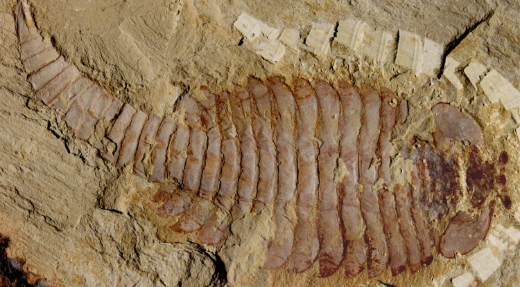
The original 520-million-year-old Fuxianhuia protensa specimen from the Chenjiang fossil beds in southwest China reveals the ancient arthropod was just shy of five inches. Credit: Photo courtesy of Xiaoya Ma, London Museum of Natural History
Science has long dictated that brains don’t fossilize, so when Nicholas Strausfeld co-authored the first ever report of a fossilized brain in a 2012 edition of Nature, it was met with “a lot of flack.”
“It was questioned by many paleontologists, who thought — and in fact some claimed in print — that maybe it was just an artifact or a one-off, implausible fossilization event,” said Strausfeld, a Regents’ professor in UA’s Department of Neuroscience.
His latest paper in Current Biology addresses these doubts head-on, with definitive evidence that, indeed, brains do fossilize.
In the paper, Strausfeld and his collaborators, including Xiaoya Ma of Yunnan Key Laboratory for Palaeobiology at China’s Yunnan University and Gregory Edgecombe of the Natural History Museum in London, analyze seven newly discovered fossils of the same species to find, in each, traces of what was undoubtedly a brain.
The species, Fuxianhuia protensa, is an extinct arthropod that roamed the seafloor about 520 million years ago. It would have looked something like a very simple shrimp. And each of the fossils — from the Chengjiang Shales, fossil-rich sites in Southwest China — revealed F. protensa’s ancient brain looked a lot like a modern crustacean’s, too.
Using scanning electron microscopy, the researchers found that the brains were preserved as flattened carbon films, which, in some fossils, were partially overlaid by tiny iron pyrite crystals. This led the research team to a convincing explanation as to how and why neural tissue fossilizes.
In another recent paper in Philosophical Transactions of the Royal Society B, Strausfeld’s experiments uncovered what it likely was about ancient environmental conditions that allowed a brain to fossilize in the first place.
The only way to become fossilized is, first, to get rapidly buried. Hungry scavengers can’t eat a carcass if it’s buried, and as long as the water is anoxic enough — that is, lacking in oxygen — a buried creature’s tissues evade consumption by bacteria as well. Strausfeld and his collaborators suspect F. protensa was buried by rapid, underwater mudslides, a scenario they experimentally recreated by burying sandworms and cockroaches in mud.
This is only step one. Step two, explained Strausfeld, is where most brains would fail: Withstanding the pressure from being rapidly buried under thick, heavy mud.
To have been able to do this, the F. protensa nervous system must have been remarkably dense. In fact, tissues of nervous systems, including brains, are densest in living arthropods. As a small, tightly packed cellular network of fats and proteins, the brain and central nervous system could pass step two, just as did the sandworm and cockroach brains in Strausfeld’s lab.
“Dewatering is different from dehydration, and it happens more gradually,” said Strausfeld, referring to the process by which pressure from the overlying mud squeezes water out of tissues. “During this process, the brain maintains its overall integrity leading to its gradual flattening and preservation. F. protensa’s tissue density appears to have made all the difference.”
Now that he and his collaborators have produced unassailable evidence that fossilized arthropod brains are more than just a one-off phenomenon, Strausfeld is working to elucidate the origin and evolution of brains over half a billion years in the past.
“People, especially scientists, make assumptions. The fun thing about science, actually, is to demolish them,” said Strausfeld.
Reference:
Xiaoya Ma, Gregory D. Edgecombe, Xianguang Hou, Tomasz Goral, Nicholas J. Strausfeld. Preservational Pathways of Corresponding Brains of a Cambrian Euarthropod. Current Biology, 2015; DOI: 10.1016/j.cub.2015.09.063
Wasp gall (Andricus kingii) attached to leaf (left) and later dissected to show inhabitant. Credit: Joyce Gross
The La Brea Tar Pits, the world’s richest Ice Age fossil site, is famous for saber-toothed cats, mammoths, and giant sloths, but it also has numerous insect and plant fossils. New research on fossil galls–abnormal plant growths caused, in this case, by tiny wasps–helps reconstruct the local habitats of Southern California at the end of the last Ice Age. The work, led by Anna R. Holden of the Richard Gilder Graduate School at the American Museum of Natural History and the La Brea Tar Pits and Museum, was recently published in the journal Quaternary Research.
“Most people associate the Ice Age with freezing temperatures and an entirely different landscape from the present,” Holden said. “But this study shows that the environment and climate around Southern California has not drastically changed since that time.”
Galls are abnormal growths on plant leaves, twigs, or branches that form in response to stimulation from invading insects, mites, bacteria, fungi, and viruses. Cynipid wasps, which lay their eggs inside plant tissue, are one of the most common gall insects. The larvae use galls for both protection and food before they emerge. Because these cynipid wasp species still live today, as is the case with most insects excavated from the tar pits, Holden, with paleobotanist Diane M. Erwin, and gall researchers Kathy Schick and Joyce Gross–all from the University of California, Berkeley–identified and linked records of the fossil gall plant hosts (mostly oaks) and their current habitat associations to the late Ice Age at the La Brea Tar Pits. This suggests that many habitats, such as coniferous forest, mixed evergreen, woodlands-savannah, and chaparral–tangled shrubs and thorny bushes–were present in Southern California between 33,000 and 44,000 years ago–the approximate date of most of the fossil galls.
“Assuming that ecological conditions required by native plants in California during the Late Pleistocene were the same as those prevailing today, the diversity of the fossil galls indicates that almost every kind of habitat existing in California at present also existed during the Late Pleistocene,” Holden said. “This suggests that a radical change or turnover in climate did not occur. Our snapshot of the prehistoric conditions of southern California is becoming clearer as we compile information from insect and plant studies.”
In order to identify certain specimens, the researchers used micro-computed tomography (CT) to compare the locations of larval chambers in modern specimens to those in the fossils.
The 13 fossil specimens the researchers studied are housed at the La Brea Tar Pits and Museum, and were recovered from different tar pits, including the Museum’s ongoing excavation “Project 23.” Dating back to the beginning of the 20th century, scientists have unearthed more than 5 million fossils representing over 600 species of plants and animals from the Tar Pits–evidence that Los Angeles was densely populated by wildlife for more than 50,000 years.
“The Tar Pits offer a unique perspective for understanding the environmental evolution of southern California,” said Luis Chiappe, vice president of research and collections at the Natural History Museum of Los Angeles County. “Saber-toothed cats speak of a bygone world while countless microfossils shed light on our changing world. In a sense, the Tar Pits are like the Roman god Janus; one face looks into the past and the other looks into the future.”
Reference:
Anna R. Holdena, Diane M. Erwind, Katherine N. Schicke, Joyce Gross. Late Pleistocene galls from the La Brea Tar Pits and their implications for cynipine wasp and native plant distribution in southern California. DOI:10.1016/j.yqres.2015.09.008
This is an image of Namacalathus hermanastes. Credit: J. Sibbick
The first animals to have complex skeletons existed about 550 million years ago, fossils of a tiny marine creature unearthed in Namibia suggest.
The find is the first to suggest the earliest complex animals on Earth — which may be related to many of today’s animal species — lived millions of years earlier than was previously known.
Until now, the oldest evidence of complex animals — which succeeded more primitive creatures that often resembled sponges or coral — came from the Cambrian Period, which began around 541 million years ago. Scientists had long suspected that complex animals had existed before then but, until now, they had no proof.
Genetic family tree data suggested that complex animals — known as bilaterians — evolved prior to the Cambrian Period. The finding suggests that bilaterians may have lived as early as 550 million years ago, during the late Ediacaran Period.
The study suggests that complex animals existed long before a period in the planet’s history — known as the Cambrian explosion — during which most major animal groups evolved.
The team studied fossils of an extinct marine animal — known as Namacalathus hermanastes — which was widespread during the Ediacaran Period. The fossils are remarkably well preserved and reveal that the species possessed a rigid skeleton made of calcium carbonate — a hard material from which the shells of marine animals are made. The complex skeletal structures are similar to those of living creatures that dwell at the bottom of the sea, the team says.
The study, published in the journal Proceedings of the Royal Society B, was funded by the Natural Environment Research Council. The research was carried out in collaboration with Lomonosov Moscow State University.
Professor Rachel Wood, of the University of Edinburgh’s School of GeoSciences, who led the study, said: “This fossil has been known for a long time, and was assumed to have been a primitive animal, such as a sponge or coral. This study suggests that it was, in fact, more advanced. We have suspected that these complex animals were present in the Ediacaran, but this study provides the first proof.”
Reference:
A. Yu. Zhuravlev, R. A. Wood, A. M. Penny. Ediacaran skeletal metazoan interpreted as a lophophorate. Proceedings of the Royal Society B: Biological Sciences, 2015; 282 (1818): 20151860 DOI: 10.1098/rspb.2015.1860
This undated image provided by Robert DePalma, shows a sketch drawn by DePalma, curator of vertebrate paleontology at the Palm Beach Museum of Natural History, which depicts the newly-discovered species of raptor called Dakotaraptor. The fossils were unearthed from the Hell Creek Formation in northwestern South Dakota. DePalma and his research team including University of Kansas paleontologists announced the new species in a study published Oct. 30, 2015, by the University of Kansas Paleontological Institute. Credit: Robert DePalma via AP
Tyrannosaurus rex may have been known as the big guy around the Hell Creek Formation 66 million years ago, but a newly discovered species of raptor would have roamed the region as one of its most lethal predators.
Dakotaraptor stood 6 feet tall at the hips yet moved like a springy, agile sprinter, reaching 30 to 40 mph rivaling today’s ostrich. But potential prey caught admiring the 17-foot-long creature’s grace stood little chance, as the strong-muscled winged Dromaeosaur boasted a vicious 9½-inch-long killing claw that could make mincemeat out of any herbivore caught in its path, said Robert DePalma, curator of vertebrate paleontology at the Palm Beach Museum of Natural History.
“It had one of the strongest killing strokes in that slashing claw of any raptor known,” DePalma said.
DePalma and his research team including University of Kansas paleontologists announced the new species in a study published Oct. 30 by the University of Kansas Paleontological Institute. Dakotaraptor helps fill a gap in body size distribution between the small bird-like Maniraptora creatures and the giant T. rex found in Hell Creek, which spans parts of northwestern South Dakota, southwestern North Dakota, eastern Montana and eastern Wyoming.
The newly discovered species roamed the earth alongside T. rex, the three-horned Triceratops and the duck-billed Edmontosaurus.
Fierce foot claw of a newly discovered species of raptor called Dakotaraptor in West Palm Beach, Fla. Credit: Kylie Ruble/Robert DePalma via AP
“Dakotaraptor coexisted with all of our favorites from our childhoods,” DePalma said. “We had no idea that such a cool and lethal creature existed right alongside them. And it was in the ground the whole time. It’s amazing.”
Thomas Holtz Jr., a senior vertebrate paleontology lecturer at the University of Maryland, said most of the raptor bones and teeth found in Hell Creek have been from small-form creatures.
“That is what is important about this find,” Holtz said. “In fact, it was rather bigger than most of us expected, almost the size of the largest known Dromaeosaurid, the much earlier Utahraptor.”
Dakotaraptor stands about as tall as Utahraptor, a species discovered in the 1990s in east-central Utah, but the raptors have completely different builds. The stockier Utahraptor, which lived about 60 million years earlier than Dakotaraptor, was an ambush predator with thicker bones and leg proportions that limited its speed, making it the “beefly bulldog of raptors,” DePalma said.
Dakotaraptor did not fly, which makes the presence of quill knobs on its arms so interesting to DePalma and other dinosaur experts. The bumps serve as reinforcement points for long wing feathers, marking the first concrete evidence that large raptors had wings.
“It really would have made this like a turkey from hell,” he said.
The feathers were clearly not just for show, and they could have been used by the dinosaur to intimidate other predators, shield its young or as a tactical method to corral prey. They might also indicate that the species evolved from a lineage that once could fly or was evolving toward flight, DePalma said.
Dakotaraptor’s leg bones, wing portions, tail vertebrae, teeth and wishbone were unearthed in 2005 from a remote area of badlands in South Dakota’s Harding County.
To find so many fossils from a single creature is extraordinary in the Hell Creek, said Peter Larson, a Black Hills-based dinosaur expert who co-authored the paper.
Hell Creek’s sediment gathered slowly over time, so most dinosaurs were eaten or carried away by other creatures before they could be buried. Many of Hell Creek’s finds consist of small individual fossils or a bone bed featuring loads of random, hard-to-match fossils, he said.
“Hell Creek is very hard to give up its secrets,” said Larson, president of the Black Hills Institute of Geological Research. “We very seldom find articulated or even associated remains.”
It’s even more difficult to find raptor remains, DePalma said, as the bird-like species’ lightweight, hollow and thin-walled bones tend to break before their preservation.
“People actually have been finding them for years without realizing it,” he said.
Dr. Julien Louys holds the jaw bone of a giant rat species discovered on East Timor, up against a comparison with the same bone of a modern rat Credit: Stuart Hay, ANU
Archaeologists with The Australian National University (ANU) have discovered fossils of seven giant rat species on East Timor, with the largest up to 10 times the size of modern rats.
Dr Julien Louys of the ANU School of Culture, History and Language, who is helping lead the project said these are the largest known rats to have ever existed.
“They are what you would call mega-fauna. The biggest one is about five kilos, the size of a small dog,” Dr Louys said.
“Just to put that in perspective, a large modern rat would be about half a kilo.”
The work is part of the From Sunda to Sahul project which is looking at the earliest human movement through Southeast Asia. Researchers are now trying to work out exactly what caused the rats to die out.
Dr Louys said the earliest records of humans on East Timor date to around 46,000 years ago, and they lived with the rats for thousands of years.
“We know they’re eating the giant rats because we have found bones with cut and burn marks,” he said.
“The funny thing is that they are co-existing up until about a thousand years ago. The reason we think they became extinct is because that was when metal tools started to be introduced in Timor, people could start to clear forests at a much larger scale.”
Dr Louys said the project team is hoping to get an idea of when humans first moved through the islands of Southeast Asia, how they were doing it and what impact they had on the ecosystems. The information can then be used to inform modern conservation efforts.
“We’re trying to find the earliest human records as well as what was there before humans arrived,” Dr Louys said.
“Once we know what was there before humans got there, we see what type of impact they had.”
Dr Louys returned from the project’s latest expedition to East Timor in August and has presented the findings at the Meetings of the Society of Vertebrate Palaeontology in Texas.
It’s an unusual job, but someone’s gotta do it. Cassandra Klos is the latest Mars Desert Research Station artist-in-residence.
Klos’ project ‘Mars On Earth’ documents engineers as they reestablish The GreenHab, a greenhouse-like Mars simulator. For Polarr, Emily von Hoffmann spoke with Klos about her work.
Emily von Hoffmann: Can you share some themes you hope to explore with this project? Cassandra Klos: Mars On Earth is about our innate need for exploration, in particular space exploration. It’s an examination of Mars research and what we’re doing on Earth right now to better equip ourselves to reach these far off places. I’m not only interested with our scientific efforts to leave Earth, but the facade we create to test these scientific endeavors. This usually comes in the form of terrestrial analog sites and simulations, but there are also many other venues that I plan to explore to fill in the gaps of the narrative.
Road to Earth, 2015. Image courtesy of Cassandra Klos.
CK: By using our planet as a sort of “stage” to play “pretend Mars” on, it kind of creates a dual reality — one where we’re off in deep space, and one where we’re still on Earth surrounded by all the comforts we know. When the Curiosity rover has an issue on Mars, there is a duplicate rover at NASA’s Jet Propulsion Laboratory to troubleshoot it. When a person is invested in a Mars analog, there is that suspension of disbelief that they are actually on Mars. It’s a trick of the mind, and I’m very fascinated in that experience!
EvH: What exactly is a terrestrial analog site? CK: A terrestrial analog site is a landscape on Earth that is geological similar to one on another planet, moon, or any other celestial body. Usually these places are the extremes of our planet, either their really cold, or really dry, or really uninhabitable areas such as volcanoes, or probably a combination. These parts of the Earth tend to give us the best understand of how our planet was created.
EvH: How did you become interested in telling a story about space? CK: As a kid I was always interested in space and astronomy, but I was (and still am) terrible at math. For a long time I was torn between going in the science direction and going in an art direction. I ended up sticking with art. By being a photographer, though, I feel like my camera allows me to explore anything I find interest in. My childhood ambitions started to funnel back into my work soon after.
EvH: During your time at the HI-SEAS analog site in Hawaii, you interviewed a team just after they’d had an eight month simulation. Can you share any favorite stories or impressions from those interviews? CK: When they were first allowed to come out of the Hab after eight months, several crew members remarked about how they felt unsure about coming out without their space suits and helmets. Of course all of us standing outside in sweatshirts and jeans laughed at the thought — but really, after eight months of not feeling fresh air and pretending that the environment around me would kill me, I think I would have a similar paranoia!
EvH: Can you tell us about your previous project, The Abductees, and how your style has grown or changed for your approach to the Mars project? (Using a camera to create a different reality, vs. exploring an attitude towards space, for instance?) CK: The Abductees is a photography project surrounding the 1961 Betty and Barney Hill alien abduction case. This story happened in my home-state of New Hampshire, so I was very close to the locations and history involved. For highly-reputable citizens Betty and Barney, the publicity of this event ruined them in many ways. Many believed they were crazy, and were shunned in their community because of what they believed, despite a plethora of documents, validating reports and sightings, and even a US Air Force report. I wanted to create the imagery that the Hills’ had no proof of. I wanted to weave a narrative that nobody could dispute.
Much of the time constructing The Abductees felt like following a movie script and executing scenes that would push the story forward. When it comes to Mars on Earth, there is a lot more freedom and allowances for spontaneity. It’s much more a of a collaboration between scientists and artists. It allows for multiple stories and perspectives. I guess I really like the lack of rigidity there is to the project right now.
EvH: Do you have a lot of thoughts about The Martian movie and the recent glut of space epics? Have they (and the sort of Kubrickian ~space chill~ they have in common) affected your aesthetic at all? CK: Oh, I have loved all the space epics lately. I just saw The Martian last weekend I thoroughly enjoyed it (scientific inconsistencies aside). And I wept during Interstellar. Just the magnitude of imagery and excitement that these movies create about future space travel is really inspiring. I don’t think they have really affected my personal aesthetic, but I wouldn’t be surprised if we’re all pulling from the same iconography.
EvH: You are the new artist-in-residence for the Mars Desert Research station in Utah, which sounds like a pretty unique job. Can you share any plans you have for your time there? CK: I’m really excited for the opportunity at the Mars Desert Research Station! There’s only been 5–6 artists-in-residence… so yeah, I’m honored to be added to that group. My crew is a bunch of engineers, so we’ll be doing a lot of maintenance on the site. We will also be reestablishing the GreenHab (the Mars greenhouse).
Image courtesy of Cassandra Klos.
I hope to explore the observatory, interview my crew members, photograph a TON, cook meals with freeze-dried ingredients, map the surrounding terrain, sketch and reflect, and be as helpful as I can be. I want to absorb as many experiences as I possibly can.
EvH: Can you share any storytellers or artists in any medium who inspire you, or whom you’re particularly enjoying right now? CK: I am inspired by contemporary artists such as Joan Fontcuberta, Trevor Paglen, Richard Mosse, An-My Le, Laura McPhee, Sophie Calle, Claire Beckett, Todd Hido, and Angela Strassheim.
Lately, I have also been looking at the photographs of the Antarctica explorer Frank Hurley. And you know, another inspiration lately has been the Apollo Archive Project Flickr account devoted to newly released film scans from the Apollo space missions!
Video footage showed a landslide in Hunza Valley, northern Pakistan after a massive earthquake hit on Monday (26 October 2015). The quake struck northern Pakistan and Afghanistan, killing at least 300 people, with at least 266 dead in Pakistan.
Monday’s initial quake of magnitude 7.5 was followed by seven aftershocks, with intensity as high as 4.8, the US Geological Survey said.
A Pacific storm swept across Southland mountains and desert areas Thursday (15 oct 2015) afternoon, unleashing intense showers that sent flows of mud and debris into homes and vehicles while cutting off the main artery between Los Angeles and the Central Valley.
A catastrophic 1982 rainstorm triggered 18,000 landslides in the Bay Area, claiming 25 lives and causing $66 million in property damage
The combination of steep slopes, weak rocks, and intense winter storms make Bay Area uplands an ideal setting for landslides
Landslides include both swift, potentially deadly debris flows and slower, but destructive deepseated slides
Learn what USGS scientists have discovered about landslide dynamics and which slopes are most susceptible to sliding
Hear the devastating stories of Bay Area residents affected by landslides and learn to recognize the danger signs
Producer/Director/Editor KAREN ADAMS Principal Videographer DOUGLAS DEVORE Graphics Animator/Artist BRYAN COLEMAN Lead Associate Editor WENDY VAN WAZER Chief Scientific Advisor RAYMOND WILSON Original Musical Score and Sound Design by DAN CANTRELL Cover Art by NANCY BROWN
The BBC has admitted that footage of a volcanic eruption screened as part of its natural history blockbuster series, Patagonia: Earth’s Secret Paradise, was faked. The scene, purporting to depict a single volcano in eruption, was actually created by splicing together eruptions from two separate volcanoes. One eruption took place in 2011, the other in 2015.
The admission is likely to trigger a new row over the use of digital techniques to make documentaries more dramatic and popular, and will embroil the corporation in further controversy at a time when its finances and remit are under parliamentary scrutiny.
Staff at the BBC’s natural history unit – which made the series – were also said to be angry about the inclusion of the doctored scene, which they fear could erode trust in their output. “If we falsify one scene, who is going to believe anything else in that programme or, for that matter, any other film that we produce,” one staff member told the Observer.
The new row comes after a bitter battle in 2011 over the BBC’s natural history series Frozen Planet, which contained footage that was said to show polar bears being born in the wild when, in fact, it was filmed at a manmade den in a German animal park.
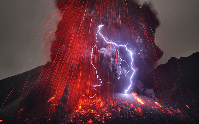
Sakurajima Volcano with Lightning Credit & Copyright: Martin Rietze/NASA
Supervolcanoes, massive eruptions with potential global consequences, appear not to follow the conventional volcano mechanics of internal pressure building until the volcano blows. Instead, a new study finds, such massive magma chambers might erupt when the roof above them cracks or collapses.
Knowledge of triggering mechanisms is crucial for monitoring supervolcano systems, including ones that lie beneath Yellowstone National Park and Long Valley, California, according to the study led by Patricia Gregg, University of Illinois professor of geology, in collaboration with professor Eric Grosfils of Pomona College and professor Shan de Silva of Oregon State University. The study was published in the Journal of Volcanology and Geothermal Research. Gregg also presented the findings this week at the annual meeting of the Geological Society of America.
“If we want to monitor supervolcanoes to determine if one is progressing toward eruption, we need better understanding of what triggers a supereruption,” Gregg said. “It’s very likely that supereruptions must be triggered by an external mechanism and not an internal mechanism, which makes them very different from the typical, smaller volcanoes that we monitor.”
A supervolcano is classed as more than 500 cubic kilometers of erupted magma volume. For comparison, Gregg said, Mount St. Helen’s ejected about one cubic kilometer of material, so a supervolcano is more than five hundred times larger.
“A typical volcano, when it erupts, can have lasting impacts across the globe,” Gregg said. “We’ve seen that in Iceland when we’ve had large ash eruptions that have completely disrupted air traffic across Europe. A supereruption takes that to the nth degree.”
The new study’s findings are contrary to a pair of papers published in the journal Nature Geoscience in 2014 that claim a link between eruption likelihood and magma buoyancy. The magma byouancy hypothesis suggested that magma may be less dense than the rock surrounding it and therefore could push up against the roof, like an ice cube bobbing in water, increasing the pressure within the chamber and triggering an eruption.
“Typically, when we think about how a volcanic eruption is triggered, we are taught that the pressure in the magma chamber increases until it causes an explosion and the volcano erupts,” Gregg said. “This is the prevailing hypothesis for how eruptions are triggered. At supervolcanic sites, however, we don’t see a lot of evidence for pressurization. When I incorporated buoyancy into my numerical models, I couldn’t reproduce the 2014 studies.”
Gregg’s numerical model incorporates all of the physics — conserving mass, energy and momentum — to calculate what would happen if a large buoyant magma body were to form in the shallow crust. The model showed that even when the chamber was huge and the difference in density was very large between the magma and he surrounding rock — an unlikely scenario — buoyancy added very little pressure to the system.
“The fact that my numerical model was not agreeing with their analytical solution suggested that there was something missing from the analytical solution. So that prompted me to look closer,” Gregg said. “What they miss in the buoyancy model is Newtonian physics: The magma may push up, but the roof pushes back down.”
The new study found that the size of the magma chamber is a much greater factor in generating supervolcanic eruptions. The buoyancy studies suggested that this correlation was due to having more material pushing up, but the Illinois-led study found that the size of the chamber affects the stability of the rock containing the chamber.
“Previous studies have found that as a magma chamber expands, it pushes the roof up and forms faults,” Gregg said. “As these very large magma chambers grow, the roof above may become unstable and it becomes easier to trigger an eruption through faulting or failure within the rock. ”
According to the model, if a crack or fault in the roof penetrates the magma chamber, the magma uses the crack as a vent to shoot to the surface. This could trigger a chain reaction that “unzips” the whole supervolcano.
Next, Gregg’s group hopes to take advantage of the advanced computing facilities available at the University of Illinois, such as the Blue Waters supercomputer at the National Center for Supercomputing Applications. The researchers are working to create 4-D models that track the evolution of the Long Valley magma chamber over time in greater detail.
“If we see a correlation between magma chamber size and the ability to erupt, it is important to know if supervolcano eruptions are triggered by internal factors or by foundering and faulting in the roof. It may mean that we have to monitor these volcanoes differently,” Gregg said. “If the trigger is an external force, whether it be an earthquake or a fault, then we should look at seismicity, what types of faults are being developed, what is the stability of the roof, and what kinds of activities are happening on the surface that could cause faulting.”
The National Science Foundation supported this work.
Reference:
Patricia M. Gregg, Eric B. Grosfils, Shanaka L. de Silva. Catastrophic caldera-forming eruptions II: The subordinate role of magma buoyancy as an eruption trigger. Journal of Volcanology and Geothermal Research, 2015; 305: 100 DOI: 10.1016/j.jvolgeores.2015.09.022
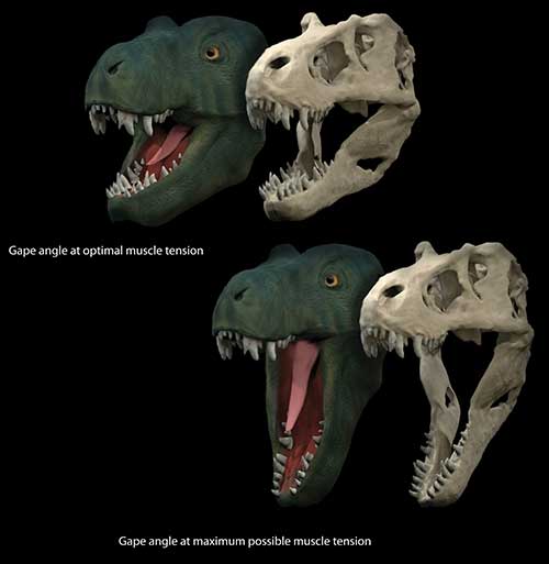
Life reconstruction and skull model of Tyrannosaurus rex showing the jaw gape at optimal position to produce muscle force and the maximal possible jaw gape Credit: Stephan Lautenschlager
Just how bad was T. rex’s bite? New research from the University of Bristol has found that the feeding style and dietary preferences of dinosaurs was closely linked to how wide they could open their jaws.
Using digital models and computer analyses, Dr Stephan Lautenschlager from Bristol’s School of Earth Sciences studied the muscle strain during jaw opening of three different theropod dinosaurs with different dietary habits. Theropods (from the Greek for “beast-footed”) were a diverse group of two-legged dinosaurs that included the largest carnivores ever to walk Earth.
Dr Lautenschlager said: “Theropod dinosaurs, such a Tyrannosaurus rex or Allosaurus, are often depicted with widely-opened jaws, presumably to emphasise their carnivorous nature. Yet, up to now, no studies have actually focused on the relation between jaw musculature, feeding style and the maximal possible jaw gape.”
The research looked at Tyrannosaurus rex, a large-sized meat-eating theropod with a massively built skull and up to 15cm long teeth; Allosaurus fragilis, a more lightly built but predatory and meat-eating theropod; and Erlikosaurus andrewsi, a closely related but plant-eating member of the theropod family.
Dr Lautenschlager said: “All muscles, including those used for closing and opening the jaw, can only stretch a certain amount before they tear. This considerably limits how wide an animal can open its jaws and therefore how and on what it can feed.”
In order to fully understand the relation between muscle strain and jaw gape, detailed computer models were created to simulate jaw opening and closing, while measuring the length changes in the digital muscles. The dinosaur species in the study were also compared to their living relatives, crocodiles and birds, for which muscle strain and maximal jaw gape are known.
The study found that the carnivorous Tyrannosaurus and Allosaurus were capable of a wide gape (up to 90 degrees), while the herbivorous Erlikosaurus was limited to small gape (around 45 degrees).
Between the two carnivores, results show that Tyrannosaurus could produce a sustained muscle (and, therefore, bite) force for a wide range of jaw angles, which would be necessary for biting through meat and skin and crushing bone.
Dr Lautenschlager said: “We know from living animals that carnivores are usually capable of larger jaw gapes than herbivores, and it is interesting to see that this also appears to be the case in theropod dinosaurs.”
The research is published in Royal Society Open Science.