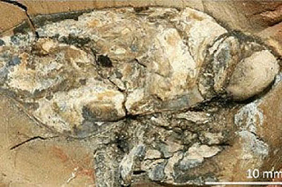
A team of researchers, among them a zoologist from the University of Cologne, has succeeded in reconstructing a 160 million year old compound eye of a fossil crustacean found in southeastern France visible. With the reconstruction of the eye, the scientists succeeded in making the structure of soft tissue visible — which was long considered to be impossible.
Zoologists from Cologne and Lyon have succeeded in discovering the internal structure of an approximately 160 million year old compound eye (Dollocaris ingens van Straelen, 1923, Thylacocephala) from the Middle Jurassic period. It was discovered at the La Voulte deposit in southeastern France. The eyes of this crustacean consist of approximately 18,000 facets, and because each of these facets contributes to the entire image as pixels contribute to a computer graphic, the eye of this crustacean belongs to the most accurate in the arthropod realm.
With the reconstruction of the eye’s structure, the scientists succeeded in making the structure of soft tissue visible — which was long considered to be impossible. Together with the palaeontologist Jean Vannier (CNRS/Université Claude Bernard Lyon 1/ENS de Lyon) and other colleagues, the zoologist Brigitte Schoenemann from the University of Cologne played a leading role in this research.
The construction of the crustacean’s high-performance eye most closely resembles that of a bee or a dragonfly. Most likely it also functioned in a similar way. A physical analysis revealed that this crustacean was active during the day and lived in the light-flooded parts of the ocean. An analysis of its stomach showed that it obviously chased smaller sea organisms and fed on them.
This research work is important because up to now researchers thought that only the hard parts of an animal, such as shells or bones, could be preserved. Hence the findings of the research team on soft tissue are ground-breaking — and they describe fossil sensory cells older than those preserved in the relatively young amber. Recently, computer-tomography has revealed that even individual sensory cells can be documented. The recent research work shows for the first time that the complete structure of a compound eye can be analysed and resolved. Thus, the team was able to open up completely new perspectives — not just for the investigation of fossilised sensory systems. It also showed that in contrast to earlier opinions, the fossil record can contribute important facts to the discussion of the evolution of visual and other internal systems.
Reference:
Jean Vannier, Brigitte Schoenemann, Thomas Gillot, Sylvain Charbonnier, Euan Clarkson. Exceptional preservation of eye structure in arthropod visual predators from the Middle Jurassic. Nature Communications, 2016; 7: 10320 DOI: 10.1038/ncomms10320
Note: The above post is reprinted from materials provided by University of Cologne.


