Credit: Henning Blom
Remember dropping your milk teeth? After a lot of wiggling the tooth finally dropped out. But in your hand was only the enamel-covered crown: the entire root of the tooth had somehow disappeared. In a paper published in Nature, a team of researchers from Uppsala University and the ESRF in France apply synchrotron x-ray tomography to a tiny jawbone of a 424 million year old fossil fish in order to illuminate the origin of this strange system of tooth replacement.
Teeth are subject to a lot of wear and tear, so it makes sense to be able to replace them during the lifetime of the animal. Surprisingly, however, the teeth of the earliest jawed vertebrates were fixed to the jaw bones and could not be shed. Tooth shedding eventually evolved independently on two occasions, using two quite different processes. In sharks and rays, the fibres that anchor the tooth to the skin of the jaw dissolve and the whole tooth simply falls out. In bony fish and land vertebrates, the developing tooth becomes attached directly to the jaw bone by a special tissue known as “bone of attachment”, and when it is time for the tooth to be shed this attachment must be severed; specialized cells come in and resorb the dentine and bone of attachment until the tooth comes loose. That’s why our milk teeth loose their roots before they are shed. But when did this process evolve?
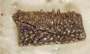
The authors of the new study decided to investigate a jaw bone of the 424 million year old fossil fish Andreolepis from Gotland in Sweden, close to the common ancestor of all living bony fish and land vertebrates. The jaw is a tiny thing, less than a centimetre in length, but it hides a wonderful secret: the internal microstructure of the bone is perfectly preserved and contains a record of its growth history. Until recently it has only been possible to see internal structures by physically cutting thin sections from the fossil and viewing them under the microscope, but this destroys the specimen and provides only a two-dimensional image that is hard to interpret.
However, at the European Synchrotron Radiation Facility (ESRF) in Grenoble, France, it is now possible to make tomographic scans that capture the same level of microscopic detail, in three dimensions, without damaging the specimen. Donglei Chen, first author of the study, has spent several years painstakingly ‘dissecting’ the scan data on the computer screen, building up a three-dimensional map of the entire sequence of tooth addition and loss – the first time an early fossil dentition has been analyzed in such detail.
“Every time a tooth was shed, the resorption process created a hollow where it had been attached. When the succeeding replacement tooth was cemented in place by bone of attachment, the old resorption surface remained as a faint buried scar within the bone tissue. I found up to four of these buried resorption surfaces under each tooth, stacked on top of each other like plates in a cupboard. This shows that the teeth were replaced again and again during the life of the fish,” explains Donglei Chen.
This is the earliest known example of tooth shedding by basal resorption, and it seems to be most similar to the process of tooth replacement seen today in primitive bony fish such as gar (Lepisosteus) and bichir (Polypterus). Like in these fish, new replacement teeth developed alongside the old ones, rather than underneath them like in us.
“The amount of biological information we get from the scans is simply astonishing. We can follow the process of growth and resorption right down to cellular level, almost like in a living animal. As we apply this technique to more early vertebrates, we will come to understand their life processes much better – and no doubt we will be in for some major surprises,” says Per Ahlberg, one of the leaders of the project.
Reference:
Donglei Chen, Henning Blom, Sophie Sanchez, Paul Tafforeau & Per E. Ahlberg (2016) The stem osteichthyan Andreolepis and the origin of tooth replacement, Nature, DOI: 10.1038/nature19812
Note: The above post is reprinted from materials provided by Uppsala University.


